You have no items in your shopping cart.
Description
Research Area
Epigenetics & Chromatin
Images & Validation
−Item 1 of 2
| Tested Applications | IHC-P, WB |
|---|---|
| Dilution Range | WB - 1:4000, IHC-P - 1:25 |
| Reactivity | Human, Mouse |
Key Properties
−| Host | Mouse |
|---|---|
| Clonality | Monoclonal |
| Isotype | IgG1,k |
| Clone No. | B035EV33X3X5 |
| Immunogen | Purified His-tagged CDH1 protein was used to produced this monoclonal antibody. |
| Target | CDH1 (HGNC:1748) |
| Molecular Weight | 97456 Da |
| Conjugation | Unconjugated |
Storage & Handling
−| Storage | Maintain refrigerated at 2-8°C for up to 2 weeks. For long term storage store at -20°C in small aliquots to prevent freeze-thaw cycles |
|---|---|
| Form/Appearance | Purified monoclonal antibody supplied in PBS with 0.09% (W/V) sodium azide. This antibody is purified through a protein G column, followed by dialysis against PBS. |
| Expiration Date | 12 months from date of receipt. |
| Disclaimer | For research use only |
Alternative Names
−Cadherin-1, CAM 120/80, Epithelial cadherin, E-cadherin, Uvomorulin, CD324, E-Cad/CTF1, E-Cad/CTF2, E-Cad/CTF3, CDH1, CDHE, UVO
Similar Products
−- Item 1 of 15
E Cadherin 1/CDH1 Rabbit Polyclonal Antibody [orb308856]
ELISA, ICC, IHC, WB
Human, Mouse, Rat
Rabbit
Polyclonal
Unconjugated
100 μg - Item 1 of 9
E Cadherin 1 CDH1 Mouse Monoclonal Antibody [orb547022]
FC, ICC, IF, IHC, WB
Human
Mouse
Monoclonal
Unconjugated
100 μg - Item 1 of 7
E cadherin Rabbit Polyclonal Antibody [orb156677]
IF, IHC-Fr, IHC-P
Rat
Human, Mouse
Rabbit
Polyclonal
Unconjugated
50 μl, 100 μl, 200 μl - Item 1 of 10
- Item 1 of 9
E-cadherin/Cdh1 Rabbit Polyclonal Antibody [orb1972562]
ELISA, FC, IF, IHC, WB
Mouse, Rat
Rabbit
Polyclonal
Unconjugated
100 μg


Quality Guarantee
Explore bioreagents carefree to elevate your research. All our products are rigorously tested for performance. If a product does not perform as described on its datasheet, our scientific support team will provide expert troubleshooting, a prompt replacement, or a refund. For full details, please see our Terms & Conditions and Buying Guide. Contact us at [email protected].
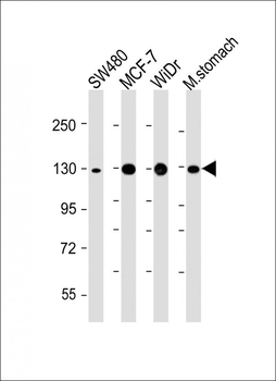
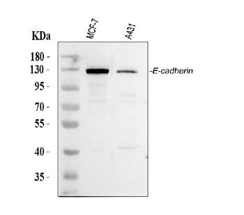
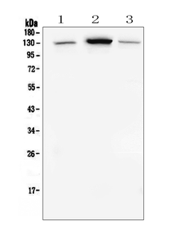
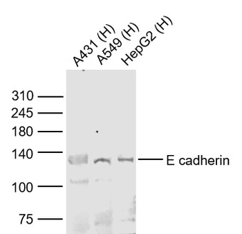
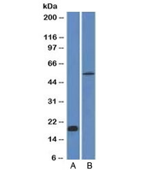
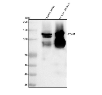
All lanes: Anti-CDH1 at 1:4000 dilution. Lane 1: SW480 whole cell lysate. Lane 2: MCF-7 whole cell lysate. Lane 3: WiDr whole cell lysate. Lane 4: Mouse stomach lysate. Lysates/proteins at 20 µg per lane. Secondary Goat Anti-mouse IgG, (H+L), Peroxidase conjugated at 1/10000 dilution. Predicted band size: 98 kDa. Blocking/Dilution buffer: 5% NFDM/TBST.
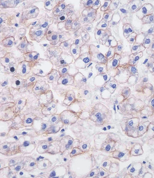
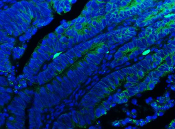
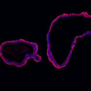
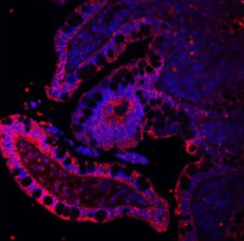
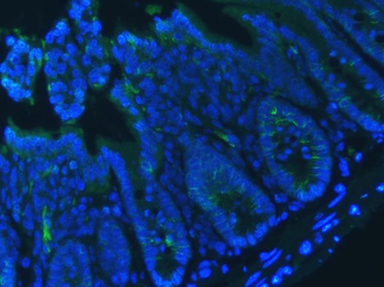
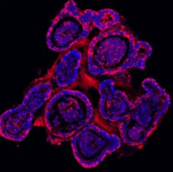
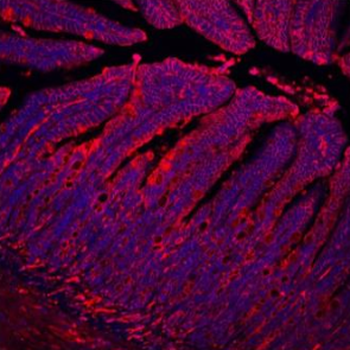
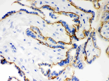
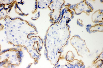
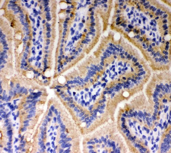
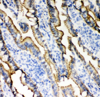
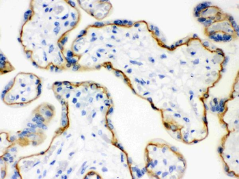
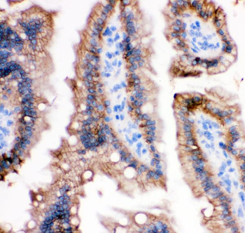
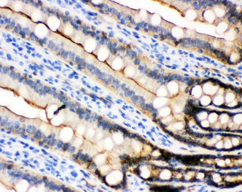
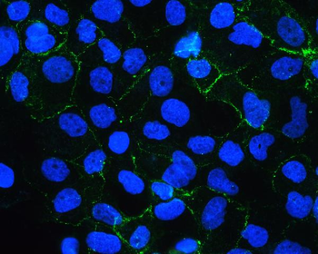
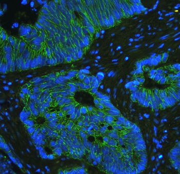
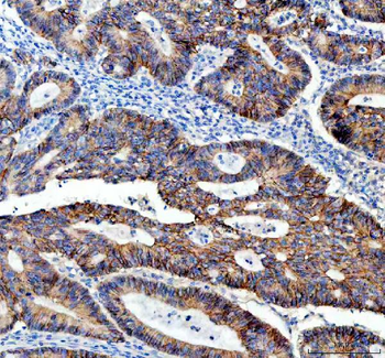
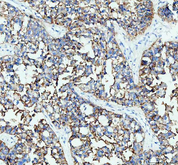
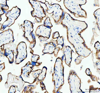
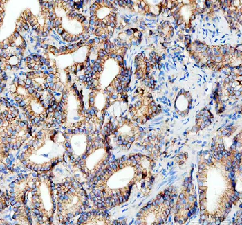
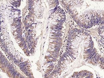
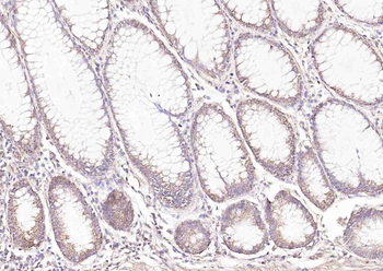
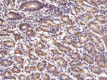
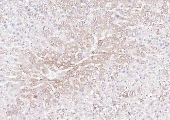
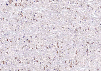
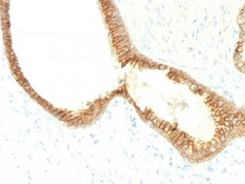
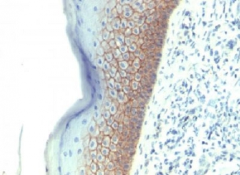
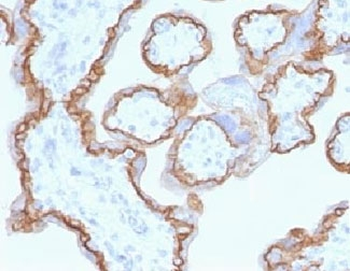
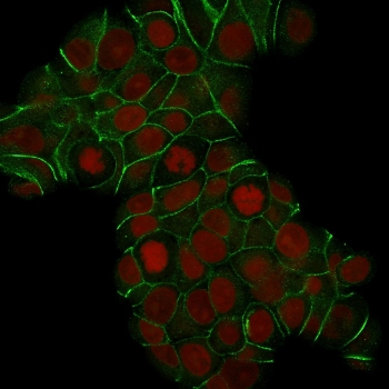
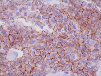
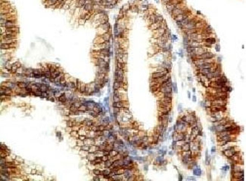
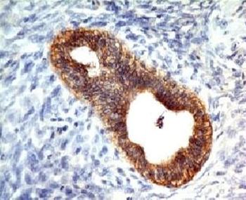
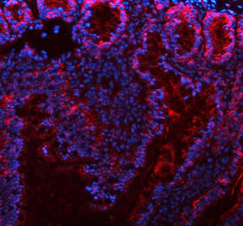
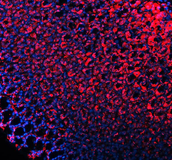
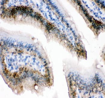
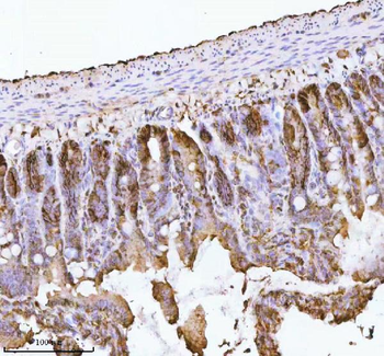
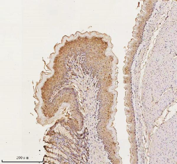
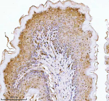
Staining CDH1 in human liver tissue sections by Immunohistochemistry (IHC-P - paraformaldehyde-fixed, paraffin-embedded sections). Tissue was fixed with formaldehyde and blocked with 3% BSA for 0.5 hour at room temperature; antigen retrieval was by heat mediation with a citrate buffer (pH6). Samples were incubated with primary antibody (1/25) for 1 hours at 37°C. A undiluted biotinylated goat polyvalent antibody was used as the secondary antibody.
Quick Database Links
UniProt Details
− No UniProt data available
NCBI Reference Sequences
−Associated Accession Numbers
Curated reference sequences for the gene transcript and protein product| Protein | NP_004351.1 |
|---|
Documents Download
Datasheet
Product Information
Request a Document
Protocol Information
WB
Western Blot (IB, immunoblot)
IHC-P
Immunohistochemistry Paraffin
CDH1 Antibody (orb1927420)

- 0.0
Based on 0 reviews
Participating in our Biorbyt product reviews program enables you to support fellow scientists by sharing your firsthand experience with our products.
Login to Submit a ReviewAvailable Sizes
Select a size below
Choose Conjugation or Carrier Free Version
Free Secondary Antibody (20 ul)0/0
Please add an antibody product to your cart first.