You have no items in your shopping cart.
Featured
Description
Research Area
Immunology & Inflammation
Images & Validation
−Item 1 of 21
| Tested Applications | ICC, IF, IHC-P, WB |
|---|---|
| Dilution Range | WB: 1:200-1000, IHC-P: 1: 100-500 |
| Reactivity | Mouse, Porcine, Rat |
Key Properties
−| Host | Rabbit |
|---|---|
| Clonality | Polyclonal |
| Isotype | IgG |
| Immunogen | KLH conjugated synthetic peptide derived from human CD41. Please contact us for the exact immunogen sequence. The peptide is available as orb374776. |
| Target | CD41 |
| Molecular Weight | 20 kDa |
| Purity | Polyclonal antibodies are purified by peptide affinity chromatography |
| Conjugation | Unconjugated |
Storage & Handling
−| Storage | Maintain refrigerated at 2-8°C for up to 2 weeks. For long term storage store at -20°C in small aliquots to prevent freeze-thaw cycles. |
|---|---|
| Form/Appearance | 10 mM PBS, 0.02% sodium azide |
| Concentration | - 100 μg (in 200 μl): 0.5 mg/ml- 200 μg (in 400 μl): 0.5 mg/ml |
| Expiration Date | 12 months from date of receipt. |
| Disclaimer | For research use only |
Alternative Names
−anti Antigen CD41 antibody, anti CD 41 antibody, anti CD41 antibody, anti CD-41 antibody, anti CD41 antigen antibody, anti CD41a antibody, anti CD41b antibody, anti GP2b antibody, anti GPalpha IIb antibody, anti GPalphaIIb antibody, anti GPIIb antibody, anti GT antibody, anti Gta antibody, anti HPA 3 antibody, anti HPA3 antibody, anti Integrin alpha 2b antibody, anti Integrin alpha IIb antibody, anti Integrin alpha IIb precursor antibody, anti Integrin alpha-IIb light chain antibody, anti ITGA 2B antibody, anti ITGA2B antibody, anti ITGAB antibody, anti Platelet fibrinogen receptor alpha antibody, anti Platelet membrane glycoprotein IIb antibody, anti Platelet specific antigen bak antibody
Similar Products
−- Item 1 of 2
CD41/ITGA2B Rabbit Polyclonal Antibody [orb184283]
FC
Bovine, Equine, Mouse, Rabbit, Rat
Human
Rabbit
Polyclonal
Unconjugated
50 μl, 100 μl, 200 μl - Item 1 of 4
CD41/Integrin alpha 2b/ITGA2B Rabbit Polyclonal Antibody [orb315155]
IHC, WB
Human, Mouse, Rat
Rabbit
Polyclonal
Unconjugated
100 μg - Item 1 of 4
CD41/Integrin Alpha 2B/ITGA2B Rabbit Polyclonal Antibody [orb1676335]
ELISA, FC, IHC, WB
Human, Rat
Rabbit
Polyclonal
Unconjugated
100 μg - Item 1 of 4
CD41 Rabbit Polyclonal Antibody [orb556881]
ICC, IHC-Fr, IHC-P, WB
Human, Mouse
Rabbit
Polyclonal
Unconjugated
100 μl - Item 1 of 2
CD41 Rabbit Polyclonal Antibody (FITC) [orb181793]
ICC, IF
Mouse, Porcine, Rat
Rabbit
Polyclonal
FITC
100 μg


Quality Guarantee
Explore bioreagents carefree to elevate your research. All our products are rigorously tested for performance. If a product does not perform as described on its datasheet, our scientific support team will provide expert troubleshooting, a prompt replacement, or a refund. For full details, please see our Terms & Conditions and Buying Guide. Contact us at [email protected].
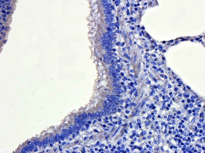
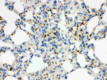
Immunohistochemical staining of pig lung tissue using anti-CD41 (dilution of primary antibody - 1:200)
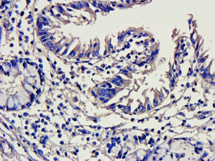
IHC-P image of pig large intestines tissue using anti-CD41 (dilution of primary antibody at 1:200)
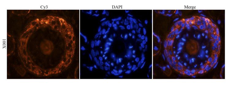
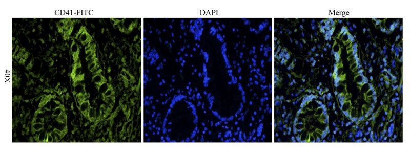
Immunofluorescence analysis of mouse skin tissue using anti-CD41 (dilution of primary antibody - 1:200)
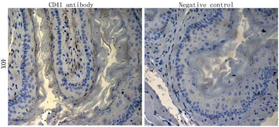
IHC-P staining of mouse skin tissue using anti-CD41 (dilution at 1:200)
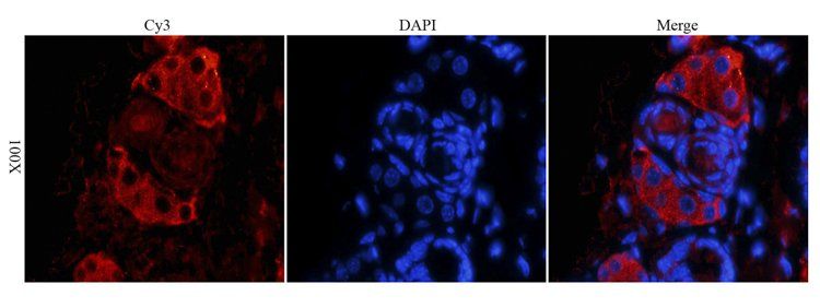
IF analysis of mouse skin tissue using CD41 antibody (dilution of primary antibody at 1:200)
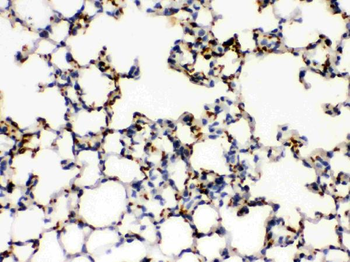
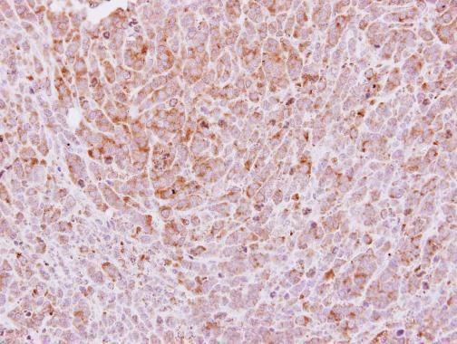
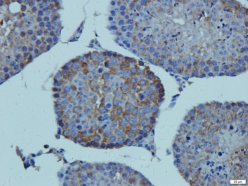
Immunohistochemical staining of paraffin embedded mouse lung tissue using CD41 antibody (primary antibody at 1:100)
IHC-P staining of mouse skin tissue using CD41 antibody (dilution at 1:200)
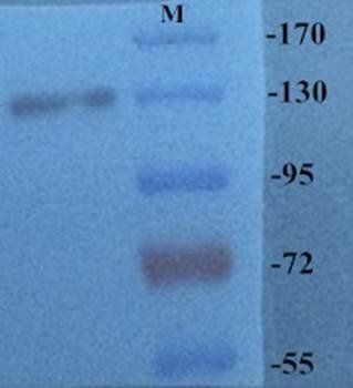
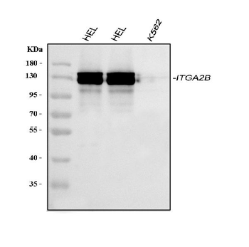
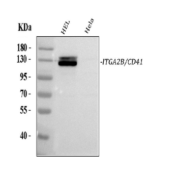
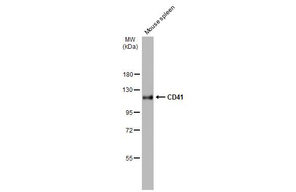
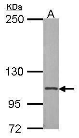
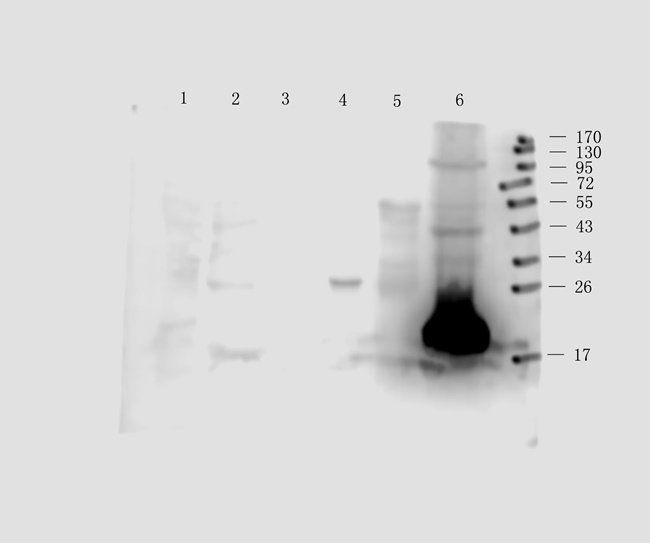
Western blot analysis of rat brain tissue using CD41 antibody (Primary antibody at 1:200)
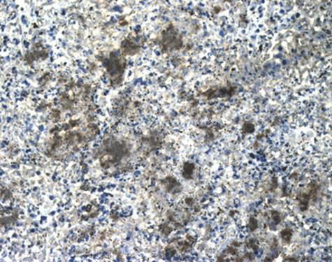
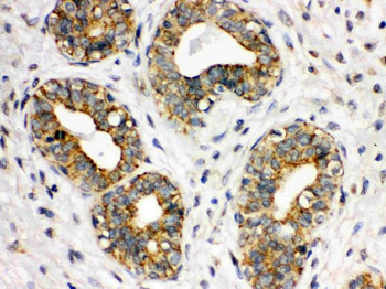
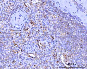
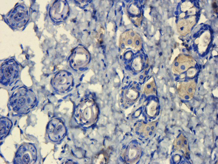
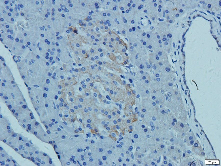
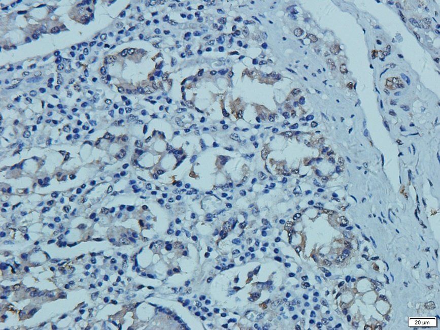
Immunohistochemical staining of paraffin embedded rat lung tissue using anti-CD41 (primary antibody at 1:100)
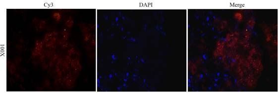
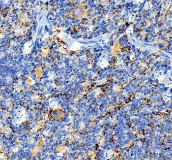
Immunofluorescence image of rat lung tissue using anti-CD41 (dilution at 1:100)
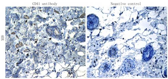
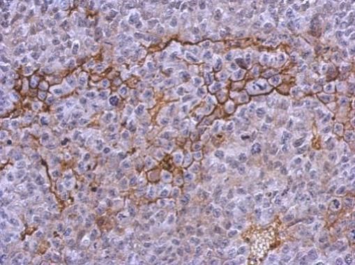
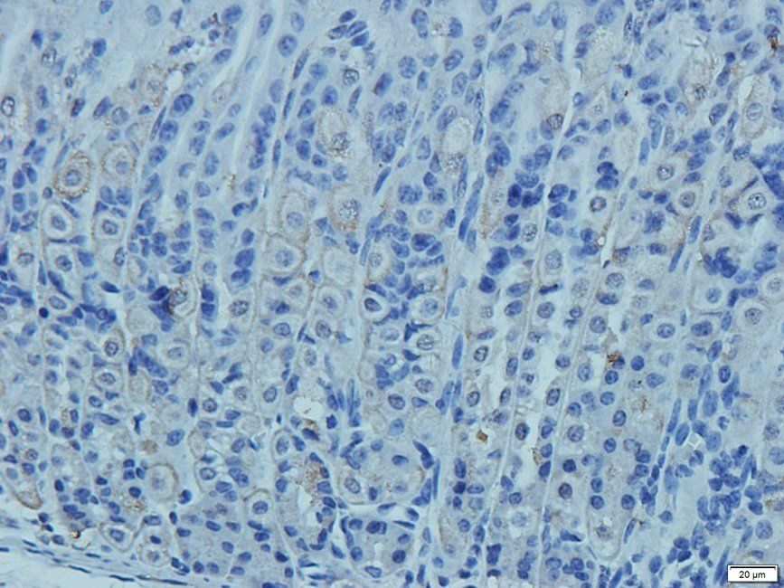
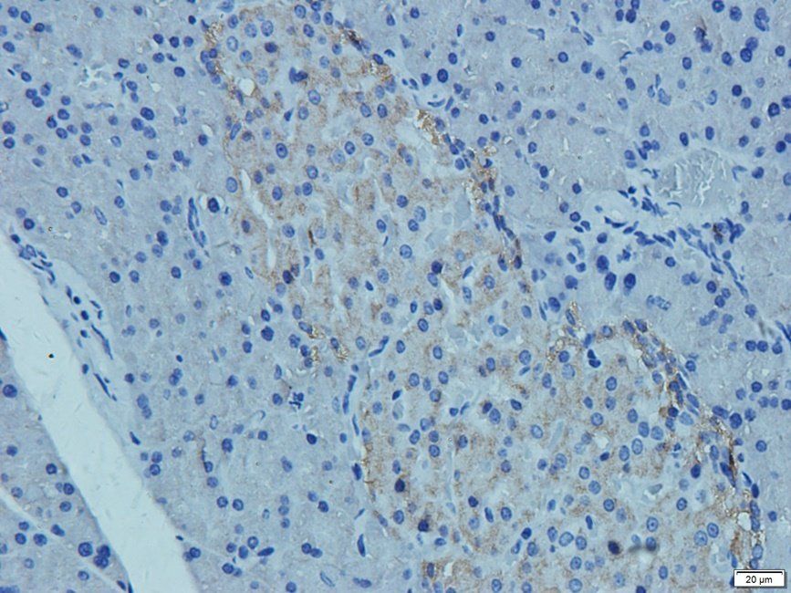
Immunohistochemical staining of rat skin tissue using CD41 antibody (dilution of primary antibody - 1:100)
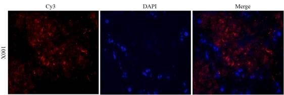
IF image of rat lung tissue using CD41 antibody (primary antibody at 1:100)
Immunohistochemical staining of rat lung tissue using anti-CD41 (dilution of primary antibody - 1:100)
Immunohistochemical staining of mouse testis tissue using Gremlin antibody (2.5 ug/ml)
Immunohistochemical staining of mouse stomach tissue using anti-Gremlin (2.5 ug/ml)
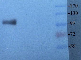
Western blot analysis of rat brain tissue using CD41 antibody (dilution of primary antibody at: 1:500)
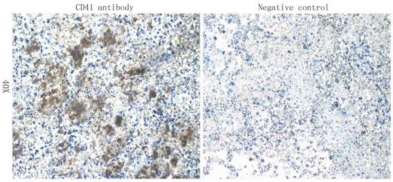
Western blot analysis of mouse stomach (lane 1), rat rectum (lane 2), mouse small intestine (lane3), rat colon (lane 4), mouse pancreas (lane 5), rat testis (lane 6) tissue using Gremlin antibody (0.5 ug/ml)
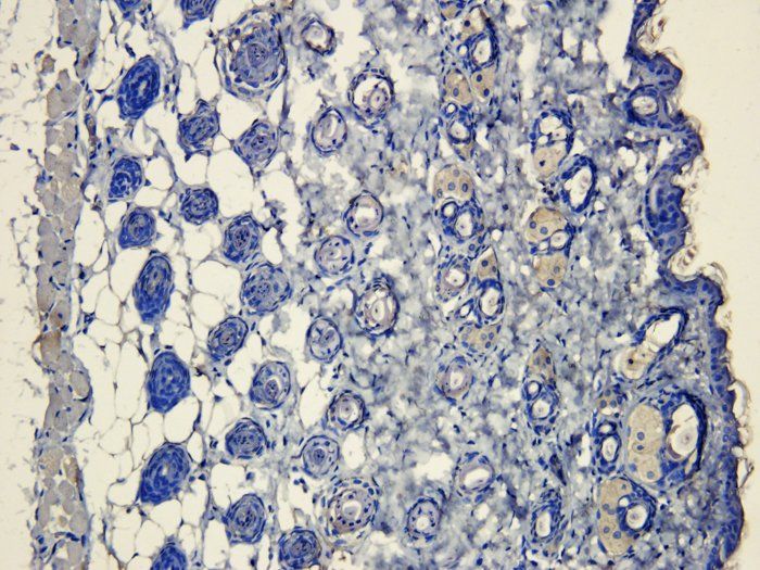
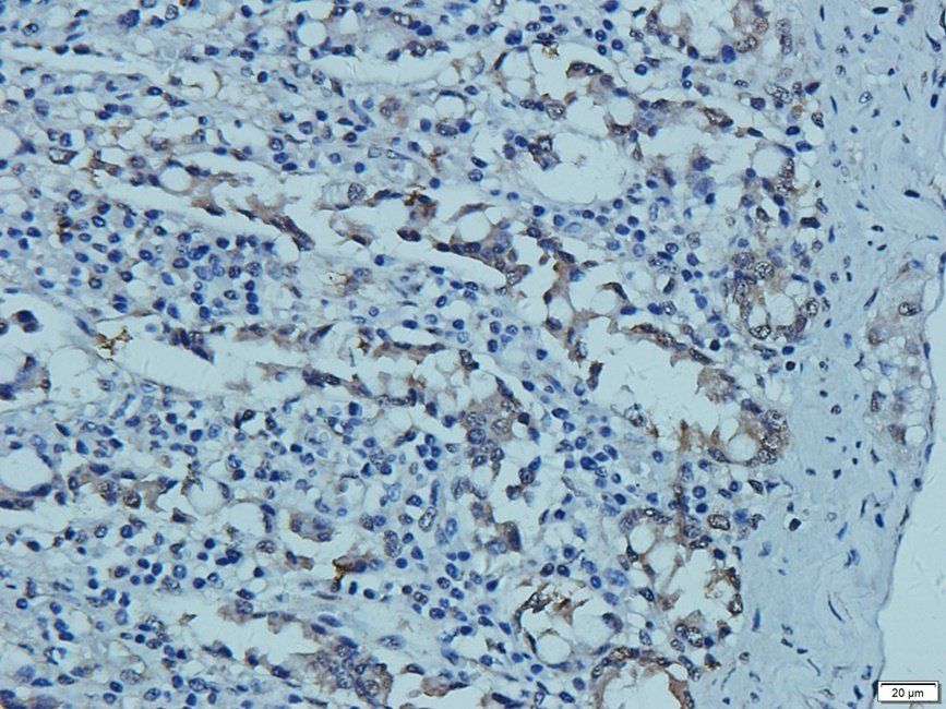
IHC-P image of rat pancreas tissue using Gremlin antibody (2.5 ug/ml)
IHC-P image of rat pancreas tissue using Gremlin antibody (2.5 ug/ml)
Immunohistochemical staining of pig small intestine tissue using anti-Gremlin (2.5 ug/ml)
IHC-P staining of pig small intestine tissue using anti-Gremlin (2.5 ug/ml)
Protocol Information
WB
Western Blot (IB, immunoblot)
IHC-P
Immunohistochemistry Paraffin
IF
Immunofluorescence
ICC
Immunocytochemistry
Filter by Applications
Filter by Species
Wei Zhang 1, Kyoko Fukazawa 2, Atsushi Mahara 2, Haiyue Jiang 3, Tetsuji Yamaoka 4 Photo-induced universal modification of small-diameter decellularized blood vessels with a hemocompatible peptide improves in vivo patency Acta Biomater, S1742-7061(24), 00012-6 (2024)
Applications
IF
Reactivity
Rat
Wei Zhang 1 2, Kyoko Fukazawa 1, Atsushi Mahara 1, Hue Thi Le 1, Raghav Soni 1, Tetsuji Yamaoka Reliable Surface Modification of ePTFE Using a Photoreactive Hemocompatible Peptide to Promote Endothelial Affinity and Antiplatelet Efficacy ACS Biomater Sci Eng, (2025)
CD41 Rabbit Polyclonal Antibody (orb4832)

- 0.0
Based on 0 reviews
Participating in our Biorbyt product reviews program enables you to support fellow scientists by sharing your firsthand experience with our products.
Login to Submit a ReviewAvailable Sizes
Select a size below
Choose Conjugation or Carrier Free Version
Free Secondary Antibody (20 ul)0/0
Please add an antibody product to your cart first.