You have no items in your shopping cart.
Featured
Description
Research Area
Neuroscience
Images & Validation
−Item 1 of 6
| Tested Applications | AM, ICC, IF, IHC, IP, WB |
|---|---|
| Dilution Range | WB (1:1000), IHC (1:1000), ICC/IF (1:100), IP (1:200) |
| Reactivity | Hamster, Human, Mouse, Rat |
| Application Notes |
Key Properties
−| Antibody Type | Recombinant Antibody |
|---|---|
| Host | Mouse |
| Clonality | Recombinant |
| Isotype | IgG1 |
| Clone No. | S57 |
| Immunogen | Fusion protein amino acids 1507-1733 (intracellular carboxyl terminus) of rabbit Cav1.2 |
| Target | Cav1.2 |
| Molecular Weight | 240kDa |
| Purification | Protein G Purified |
| Conjugation | APC |
Storage & Handling
−| Storage | Conjugated antibodies should be stored according to the product label |
|---|---|
| Buffer/Preservatives | 95.46mM Phosphate, 2.48mM MES and 2mM EDTA |
| Concentration | 1 mg/ml |
| Expiration Date | 12 months from date of receipt. |
| Disclaimer | For research use only |
Alternative Names
−Voltage-dependent L-type calcium channel subunit alpha-1C, Cav1.2, Calcium channel, L type, alpha-1 polypeptide, isoform 1, cardiac muscle, Voltage-gated calcium channel subunit alpha Cav1.2, DHPR alpha-1 subunit, CACH3, CACN4, CACNL1A2, alpha-1 subunit voltage-dependent calcium channel, calcium channel voltage-dependent L type alpha 1C subunit1, calcium channel L type, alpha 1 polypeptide isoform 1 cardiac muscle, calcium channel cardiac dihydropyridine-sensitive alpha-1 subunit, voltage-gated L-type calcium channel Cav1.2 alpha 1 subunit splice variant 10
Similar Products
−
CACNA1C Rabbit Polyclonal Antibody (APC) [orb1006665]
IF
Bovine, Canine, Equine, Human, Mouse, Porcine, Rabbit, Rat
Rabbit
Polyclonal
APC
100 μlCACNA1C Rabbit Polyclonal Antibody (APC) [orb1006635]
IF
Bovine, Canine, Equine, Human, Mouse, Porcine, Rabbit, Rat
Rabbit
Polyclonal
APC
100 μlCACNA1C Rabbit Polyclonal Antibody (APC-Cy7) [orb2434672]
IF
Bovine, Canine, Equine, Human, Mouse, Porcine, Rabbit, Rat
Rabbit
Polyclonal
APC/Cy7
100 μlCACNA1C Rabbit Polyclonal Antibody (APC-Cy5.5) [orb2434673]
IF
Bovine, Canine, Equine, Human, Mouse, Porcine, Rabbit, Rat
Rabbit
Polyclonal
APC/Cy5.5
100 μlCACNA1C Rabbit Polyclonal Antibody (APC-Cy7) [orb2434899]
IF
Bovine, Canine, Equine, Human, Mouse, Porcine, Rabbit, Rat
Rabbit
Polyclonal
APC/Cy7
100 μl
Quality Guarantee
Explore bioreagents carefree to elevate your research. All our products are rigorously tested for performance. If a product does not perform as described on its datasheet, our scientific support team will provide expert troubleshooting, a prompt replacement, or a refund. For full details, please see our Terms & Conditions and Buying Guide. Contact us at [email protected].
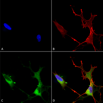
Immunocytochemistry/Immunofluorescence analysis using Mouse Anti-Cav1.2 Monoclonal Antibody, Clone S57. Tissue: Neuroblastoma cells (SH-SY5Y). Species: Human. Fixation: 4% PFA for 15 min. Primary Antibody: Mouse Anti-Cav1.2 Monoclonal Antibody at 1:50 for overnight at 4°C with slow rocking. Secondary Antibody: AlexaFluor 488 at 1:1000 for 1 hour at RT. Counterstain: Phalloidin-iFluor 647 (red) F-Actin stain; Hoechst (blue) nuclear stain at 1:800, 1.6mM for 20 min at RT. (A) Hoechst (blue) nuclear stain. (B) Phalloidin-iFluor 647 (red) F-Actin stain. (C) Cav1.2 Antibody (D) Composite.
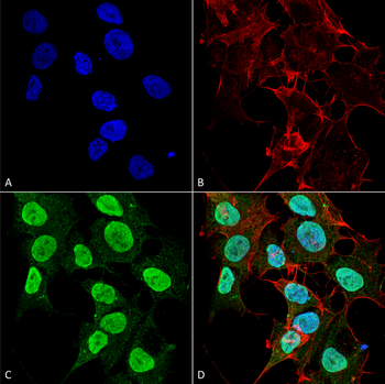
Immunocytochemistry/Immunofluorescence analysis using Mouse Anti-Cav1.2 Monoclonal Antibody, Clone S57. Tissue: Neuroblastoma cell line (SK-N-BE). Species: Human. Fixation: 4% Formaldehyde for 15 min at RT. Primary Antibody: Mouse Anti-Cav1.2 Monoclonal Antibody at 1:100 for 60 min at RT. Secondary Antibody: Goat Anti-Mouse ATTO 488 at 1:200 for 60 min at RT. Counterstain: Phalloidin Texas Red F-Actin stain; DAPI (blue) nuclear stain at 1:1000, 1:5000 for 60 min at RT, 5 min at RT. Localization: Cell Membrane, Membrane, Cytoplasm, Nucleoplasm. Magnification: 60X. (A) Phalloidin Texas Red F-Actin stain; DAPI (blue) nuclear stain. (B) Anti-Cav1.2 Antibody. (C) Composite. (A) DAPI (blue) nuclear stain. (B) Phalloidin Texas Red F-Actin stain. (C) Cav1.2 Antibody. (D) Composite.
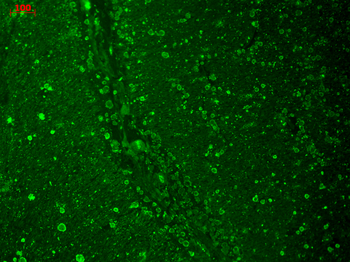
Immunohistochemistry analysis using Mouse Anti-CaV1.2 Calcium Channel Monoclonal Antibody, Clone S57. Tissue: hippocampus. Species: Human. Fixation: 10% formalin. Primary Antibody: Mouse Anti-CaV1.2 Calcium Channel Monoclonal Antibody at 1:100 for 1 hour at RT. Secondary Antibody: FITC Goat Anti-Mouse (green) at 1:50 for 1 hour at RT.
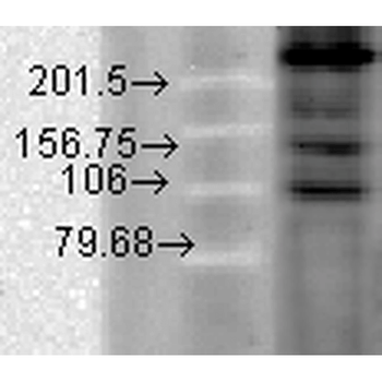
Western Blot analysis of Hamster T-CHO cell lysate showing detection of CaV1.2 Calcium Channel protein using Mouse Anti-CaV1.2 Calcium Channel Monoclonal Antibody, Clone S57. Primary Antibody: Mouse Anti-CaV1.2 Calcium Channel Monoclonal Antibody at 1:1000.
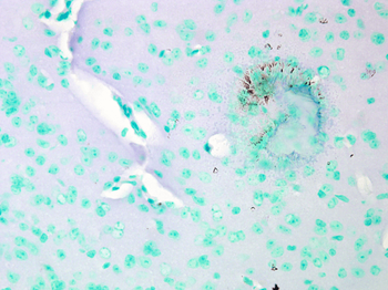
Immunohistochemistry analysis using Mouse Anti-CaV1.2 Calcium channel Monoclonal Antibody, Clone S57. Tissue: Brain Tissue. Species: Mouse. Fixation: Formalin. Primary Antibody: Mouse Anti-CaV1.2 Calcium channel Monoclonal Antibody at 1:10000 for 12 hours at 4°C. Secondary Antibody: Biotin Goat Anti-Mouse at 1:2000 for 1 hour at RT. Counterstain: Mayer Hematoxylin (purple/blue) nuclear stain at 200 μl for 2 minutes at RT. Magnification: 40x.
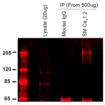
Immunoprecipitation analysis using Mouse Anti-CaV1.2 Calcium Channel Monoclonal Antibody, Clone S57. Tissue: INS-1E cells. Species: Rat. Primary Antibody: Mouse Anti-CaV1.2 Calcium Channel Monoclonal Antibody at 1:200.
Quick Database Links
UniProt Details
− No UniProt data available
NCBI Gene Details
− No NCBI Gene data available
NCBI Reference Sequences
−Associated Accession Numbers
Curated reference sequences for the gene transcript and protein product| Protein | NP_001129994.1 |
|---|
Documents Download
Datasheet
Product Information
Request a Document
Protocol Information
WB
Western Blot (IB, immunoblot)
IHC
Immunohistochemistry
IF
Immunofluorescence
ICC
Immunocytochemistry
IP
Immunoprecipitation
Cav1.2 Antibody (APC) (orb148160)

- 0.0
Based on 0 reviews
Participating in our Biorbyt product reviews program enables you to support fellow scientists by sharing your firsthand experience with our products.
Login to Submit a ReviewAvailable Sizes
Select a size below
Choose Conjugation or Carrier Free Version
Free Secondary Antibody (20 ul)0/0
Please add an antibody product to your cart first.