You have no items in your shopping cart.
Blocking Buffer For Fluorescent Western Blotting
SKU: orb348640
Description
Research Area
Metabolism Research
Images & Validation
−Item 1 of 7
| Application Notes |
|---|
Key Properties
−| Purity | Blocking buffer for Western Blotting was prepared using ultra pure reagents dissolved in pharmaceutical grade water (WFI) and consists of a proprietary protein formulation in TRIS buffered saline at pH 7.6 with thimerosal added as an antimicrobial agent. |
|---|---|
| Conjugation | Unconjugated |
Storage & Handling
−| Storage | Store vial at 4° C prior to opening. DO NOT FREEZE. |
|---|---|
| Form/Appearance | Liquid (sterile filtered) |
| Buffer/Preservatives | Preservative: Thimerosal is added as an antimicrobial agent.; Buffer: See application note. |
| Concentration | 1x |
| Expiration Date | 12 months from date of receipt. |
| Hazard Information | Non-Toxic |
| Disclaimer | For research use only |
Alternative Names
−IRDye Blocking Buffer, Fluorescent Blocking Buffer, Blocking Solution
Similar Products
−- Item 1 of 7
- Item 1 of 7
- Item 1 of 7
- Item 1 of 1
- Item 1 of 1


Quality Guarantee
Explore bioreagents carefree to elevate your research. All our products are rigorously tested for performance. If a product does not perform as described on its datasheet, our scientific support team will provide expert troubleshooting, a prompt replacement, or a refund. For full details, please see our Terms & Conditions and Buying Guide. Contact us at [email protected].
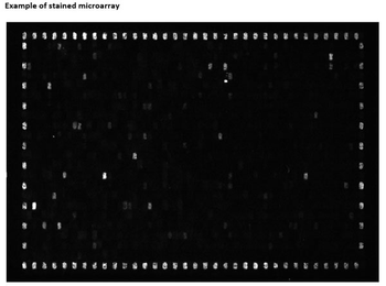
702 Peptides are printed in duplicates randomly distributed on the microarray. Control peptides (HA and FLAG controls) are located in a square surrounding the peptides of interest. As secondary antibody DyLight™ 549 conjugated goat anti-human IgG antibody and for the FLAG control peptide a mouse anti-FLAG-Cy3 antibody were used.
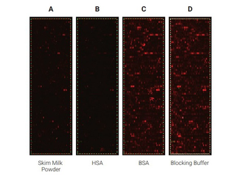
Comparison of the performance of different blocking reagents in epitope mappings with PEPperCHIP® Peptide Microarrays. The PEPperCHIP® Peptide Microarrays were blocked for 30 minutes with either 2% skim milk powder (A), 1% HSA (B), 1% BSA (C) or 100% Biorbyt Blocking Buffer [p/n orb348637] (D). A human serum sample was assayed at dilution 1:200, followed by detection with secondary goat anti-Human IgG (H+L) DyLight™ 680 Antibody and a control anti-HA (12CA5)-DyLight™ 800 Antibody. Red spots = sample IgG response and frame of polio control peptides, green spots = frame of HA control peptides.

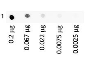
Dot Blot of Human IgA Fluorescein using orb348637. Antigen: Human IgA Fluorescein. Load: 3-fold serial dilution starting at 200 ng. Block: orb348637 for 30 min at RT.
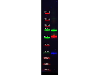
Multiplex western blot results using orb348637. Biorbyt Mouse-a-GST (orb344400, blue), Rabbit anti-Transferrin (orb750703), and Goat-anti-Alpha-1-Anti-Trypsin (orb750391) were used in a multiplex system to detect target proteins under reducing conditions in albumin depleted human serum with 320 ng of added GST. Sample was run by SDS-PAGE, transferred to 0.2 um PVDF using the BioRad Trans-Blot Turbo and blocked in 2.5% Blotto, 2.5% BSA, 0.02% Tween over night at 4°C. Membrane was probed with three primary antibodies at 1:1000 dilution (in orb348637 over night at 4°C). Detection shown was using DyLight™549 Donkey anti-Rabbit IgG (shown as green) DyLight™488 Donkey anti-Mouse IgG (shown as blue), and DyLight™649 Donkey anti-Goat IgG (shown as red) at 1:10000 (in orb348637 at 30 min RT). Blots were washed, rinsed in methanol, dried.
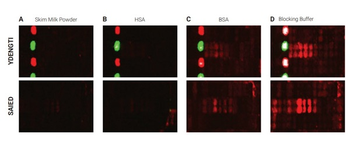
Selected sections of the PEPperCHIP® Peptide Microarrays after assay with different blocking reagents. The microarrays were blocked for 30 minutes with either 2% skim milk powder (A), 1% HSA (B), 1% BSA (C) or 100% Biorbyt Blocking Buffer [p/n orb348637] (D), respectively. A human serum sample was assayed at dilution 1:200, followed by detection with secondary goat Anti-Human IgG (H+L) DyLight™ 680 Antibody. Red spots = sample responses and polio control peptides, green spots = HA control peptides. The underlying binding motifs of the respective sections are indicated on the left.

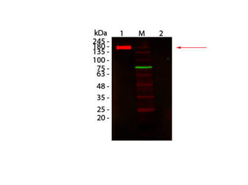
Western Blot of Fluorescent TrueBlot®: Anti-Rabbit IgG DyLight 680 Conjugated using orb348637. Lane 1: Rabbit IgG, Non-denatured. Lane 2: Rabbit IgG, Denatured. Load: 50 ng per lane. Primary antibody: none. Secondary antibody: Fluorescent TrueBlot®: Anti-Rabbit IgG DyLight 680 Conjugated antibody at 1:1000 for 60 min at RT. Block: orb348637 for 30 min at RT. Predicted: 160 kDa for non-denatured; observed: 170-180 kDa for non-denatured. Band migrates at slightly higher molecular weight.

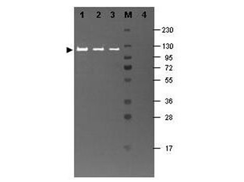
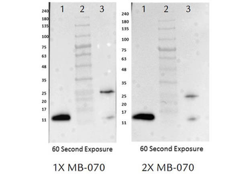
Western blot results using orb348637 and Fluorescein conjugated anti-b-Galactosidase antibody shows a band at ~117 kDa. Lanes 1 - 3 loaded with 60 ng, 30 ng and 15 ng, respectively of b-Gal present in partially purified preparations (arrowhead). Lane 4 shows no cross reactivity with proteins present in a non-specific control E.coli lysate. Proteins were resolved on a 4-20% Tris-Glycine gel by SDS-PAGE and transferred to nitrocellulose and blocking using Blocking Buffer for Fluorescent Western Blotting (p/n orb348637). The membrane was probed with fluorescein conjugated anti-b-Galactosidase (p/n orb344848) diluted to 1:10000. Reaction occurred for 2 hours at room temperature. Molecular weight estimation was made by comparison to a prestained MW marker in lane M.
Documents Download
Datasheet
Product Information
Request a Document
Blocking Buffer For Fluorescent Western Blotting (orb348640)

- 0.0
Based on 0 reviews
Participating in our Biorbyt product reviews program enables you to support fellow scientists by sharing your firsthand experience with our products.
Login to Submit a ReviewAvailable Sizes
Select a size below