You have no items in your shopping cart.
Blocking Buffer For Fluorescent Western Blotting 3-Pack
SKU: orb348638
Description
Research Area
Metabolism Research
Images & Validation
−Item 1 of 7
| Application Notes |
|---|
Key Properties
−| Purity | Blocking buffer was prepared using ultra pure reagents dissolved in pharmaceutical grade water (WFI) and consists of a proprietary protein formulation in TRIS buffered saline at pH 7.6 with thimerosal added as an antimicrobial agent. |
|---|---|
| Conjugation | Unconjugated |
Storage & Handling
−| Storage | Store blocking buffer at 4° C prior to opening. DO NOT FREEZE. |
|---|---|
| Form/Appearance | Liquid (sterile filtered) |
| Buffer/Preservatives | Preservative: Thimerosal is added as an antimicrobial agent.; Buffer: See application note. |
| Concentration | 1X |
| Expiration Date | 6 months from date of receipt. |
| Hazard Information | Non-Toxic |
| Disclaimer | For research use only |
Alternative Names
−Multiplex Blocking Buffer, Fluorescent Blocking Buffer, Blocking Solution, Blocking Buffer Western Blot, IRDye Western Blot Blocking Buffer, Alexa Dye Blocking Buffer, DyLight Blocking Buffer
Quality Guarantee
Explore bioreagents carefree to elevate your research. All our products are rigorously tested for performance. If a product does not perform as described on its datasheet, our scientific support team will provide expert troubleshooting, a prompt replacement, or a refund. For full details, please see our Terms & Conditions and Buying Guide. Contact us at [email protected].
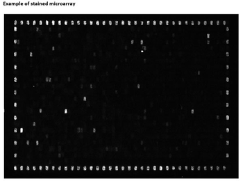
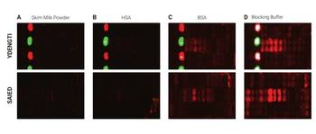
702 Peptides are printed in duplicates randomly distributed on the microarray. Control peptides (HA and FLAG controls) are located in a square surrounding the peptides of interest. As secondary antibody DyLight™ 549 conjugated goat anti-human IgG antibody and for the FLAG control peptide a mouse anti-FLAG-Cy3 antibody were used.
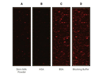
Comparison of the performance of different blocking reagents in epitope mappings with PEPperCHIP® Peptide Microarrays. The PEPperCHIP® Peptide Microarrays were blocked for 30 minutes with either 2% skim milk powder (A), 1% HSA (B), 1% BSA (C) or 100% Biorbyt Blocking Buffer [p/n orb348637] (D). A human serum sample was assayed at dilution 1:200, followed by detection with secondary goat anti-Human IgG (H+L) DyLight™ 680 Antibody and a control anti-HA (12CA5)-DyLight™ 800 Antibody. Red spots = sample IgG response and frame of polio control peptides, green spots = frame of HA control peptides.
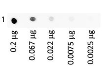
Dot Blot of Human IgA Fluorescein using orb348637. Antigen: Human IgA Fluorescein. Load: 3-fold serial dilution starting at 200 ng. Block: orb348637 for 30 min at RT.
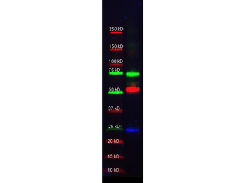
Multiplex western blot results using orb348637. Biorbyt Mouse-a-GST (orb344400, blue), Rabbit anti-Transferrin (orb750703), and Goat-anti-Alpha-1-Anti-Trypsin (orb750391) were used in a multiplex system to detect target proteins under reducing conditions in albumin depleted human serum with 320 ng of added GST. Sample was run by SDS-PAGE, transferred to 0.2 um PVDF using the BioRad Trans-Blot Turbo and blocked in 2.5% Blotto, 2.5% BSA, 0.02% Tween over night at 4°C. Membrane was probed with three primary antibodies at 1:1000 dilution (in orb348637 over night at 4°C). Detection shown was using DyLight™549 Donkey anti-Rabbit IgG (shown as green) DyLight™488 Donkey anti-Mouse IgG (shown as blue), and DyLight™649 Donkey anti-Goat IgG (shown as red) at 1:10000 (in orb348637 at 30 min RT). Blots were washed, rinsed in methanol, dried.
Selected sections of the PEPperCHIP® Peptide Microarrays after assay with different blocking reagents. The microarrays were blocked for 30 minutes with either 2% skim milk powder (A), 1% HSA (B), 1% BSA (C) or 100% Biorbyt Blocking Buffer [p/n orb348637] (D), respectively. A human serum sample was assayed at dilution 1:200, followed by detection with secondary goat Anti-Human IgG (H+L) DyLight™ 680 Antibody. Red spots = sample responses and polio control peptides, green spots = HA control peptides. The underlying binding motifs of the respective sections are indicated on the left.
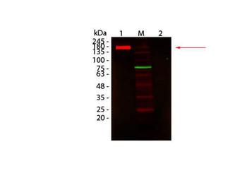
Western Blot of Fluorescent TrueBlot®: Anti-Rabbit IgG DyLight 680 Conjugated using orb348637. Lane 1: Rabbit IgG, Non-denatured. Lane 2: Rabbit IgG, Denatured. Load: 50 ng per lane. Primary antibody: none. Secondary antibody: Fluorescent TrueBlot®: Anti-Rabbit IgG DyLight 680 Conjugated antibody at 1:1000 for 60 min at RT. Block: orb348637 for 30 min at RT. Predicted: 160 kDa for non-denatured; observed: 170-180 kDa for non-denatured. Band migrates at slightly higher molecular weight.
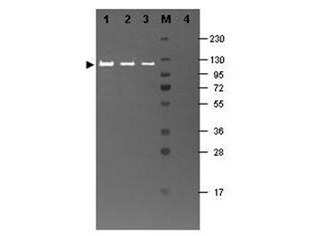
Western blot results using orb348637 and Fluorescein conjugated anti-b-Galactosidase antibody shows a band at ~117 kDa. Lanes 1 - 3 loaded with 60 ng, 30 ng and 15 ng, respectively of b-Gal present in partially purified preparations (arrowhead). Lane 4 shows no cross reactivity with proteins present in a non-specific control E.coli lysate. Proteins were resolved on a 4-20% Tris-Glycine gel by SDS-PAGE and transferred to nitrocellulose and blocking using Blocking Buffer for Fluorescent Western Blotting (p/n orb348637). The membrane was probed with fluorescein conjugated anti-b-Galactosidase (p/n orb344848) diluted to 1:10000. Reaction occurred for 2 hours at room temperature. Molecular weight estimation was made by comparison to a prestained MW marker in lane M.
Documents Download
Datasheet
Product Information
Request a Document
Blocking Buffer For Fluorescent Western Blotting 3-Pack (orb348638)

- 0.0
Based on 0 reviews
Participating in our Biorbyt product reviews program enables you to support fellow scientists by sharing your firsthand experience with our products.
Login to Submit a ReviewAvailable Sizes
Select a size below