You have no items in your shopping cart.
Featured
Description
Research Area
Cell Biology
Images & Validation
−Item 1 of 9
| Tested Applications | IF, IHC-Fr, IHC-P, WB |
|---|---|
| Dilution Range | WB=1:2000-5000, IHC-P=1:50-200, IHC-F=1:50-200, IF=1:50-200 |
| Reactivity | Human, Mouse, Rat |
| Predicted Reactivity | Mouse, Rat |
Key Properties
−| Antibody Type | Primary Antibody |
|---|---|
| Host | Rabbit |
| Clonality | Recombinant |
| Isotype | IgG |
| Clone No. | B4H33 |
| Immunogen | A synthesized peptide derived from human beta Tubulin (400-444aa) |
| Target | TUBB |
| Molecular Weight | 50 kDa |
| Purification | Affinity purified by Protein A |
| Conjugation | Unconjugated |
Storage & Handling
−| Storage | Maintain refrigerated at 2-8°C for up to 2 weeks. For long term storage store at -20°C in small aliquots to prevent freeze-thaw cycles. |
|---|---|
| Form/Appearance | Liquid |
| Buffer/Preservatives | 0.01M TBS (pH7.4) with 1% rAlbumin, 0.02% Proclin300 and 50% Glycerol. |
| Concentration | 1mg/ml |
| Expiration Date | 12 months from date of receipt. |
| Disclaimer | For research use only |
Alternative Names
−CDCBM6; CSCSC1; M40; OK/SW-cl.56; TUBB1; TUBB5; B130022C14Rik; M(beta)5; Tubb; TBB5_HUMAN; Tubulin beta-5 chain; TBB5_MOUSE; TBB5_RAT; tubulin beta class I; tubulin, beta polypeptide; tubulin, beta; tubulin, beta class I; class I beta-tubulin; beta1-tubulin
Similar Products
−- Item 1 of 9
Tubulin beta-III Recombinant Rabbit Monoclonal Antibody [orb612226]
FC, ICC, IF, IHC-Fr, IHC-P, WB
Mouse, Rat
Human, Mouse, Rat
Rabbit
Recombinant
Unconjugated
50 μl, 100 μl, 25 μl 
PtX Rabbit Anti-Beta Tubulin (S11B) Recombinant Antibody [orb1736858]
WB
Human, Mouse
Plant
Monoclonal
Unconjugated
100 μg- Item 1 of 1
Beta I Tubulin Recombinant Rabbit Monoclonal Antibody [orb2562401]
FC, ICC, IF, IHC-Fr, IHC-P, WB
Monkey
Human, Mouse, Rat
Rabbit
Recombinant
Unconjugated
50 μl, 100 μl, 25 μl - Item 1 of 1
Beta 4B/2A/2B Tubulin Recombinant Rabbit Monoclonal Antibody [orb2562928]
FC, IF, IHC-Fr, IHC-P, IP, WB
Human, Mouse, Rat
Human, Mouse, Rat
Rabbit
Recombinant
Unconjugated
50 μl, 100 μl, 25 μl Rabbit beta-Tubulin Recombinant Monoclonal Antibody [orb1519398]
FC, ICC, IHC, IP, WB
Human, Mouse
Rabbit
Recombinant
Unconjugated
100 μg (BSA-free)


Quality Guarantee
Explore bioreagents carefree to elevate your research. All our products are rigorously tested for performance. If a product does not perform as described on its datasheet, our scientific support team will provide expert troubleshooting, a prompt replacement, or a refund. For full details, please see our Terms & Conditions and Buying Guide. Contact us at [email protected].
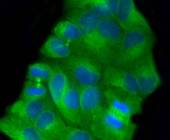
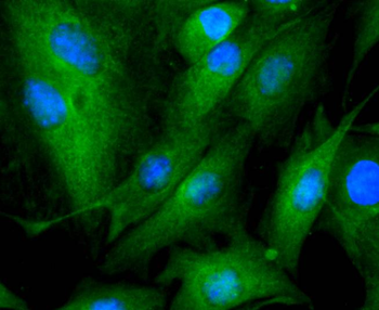
ICC staining of beta Tubulin in Hela cells (green). Formalin fixed cells were permeabilized with 0.1% Triton X-100 in TBS for 10 minutes at room temperature and blocked with 10% negative goat serum for 15 minutes at room temperature. Cells were probed with the primary antibody (orb1499401, 1/50) for 1 hour at room temperature, washed with PBS. Alexa Fluor®488 conjugate-Goat anti-Rabbit IgG was used as the secondary antibody at 1/1000 dilution. The nuclear counter stain is DAPI (blue).
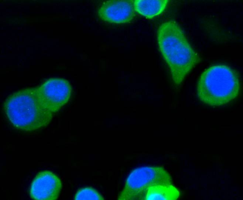
ICC staining of beta Tubulin in N2A cells (green). Formalin fixed cells were permeabilized with 0.1% Triton X-100 in TBS for 10 minutes at room temperature and blocked with 10% negative goat serum for 15 minutes at room temperature. Cells were probed with the primary antibody (orb1499401, 1/50) for 1 hour at room temperature, washed with PBS. Alexa Fluor®488 conjugate-Goat anti-Rabbit IgG was used as the secondary antibody at 1/1000 dilution. The nuclear counter stain is DAPI (blue).
ICC staining of beta Tubulin in PC-12 cells (green). Formalin fixed cells were permeabilized with 0.1% Triton X-100 in TBS for 10 minutes at room temperature and blocked with 10% negative goat serum for 15 minutes at room temperature. Cells were probed with the primary antibody (orb1499401, 1/50) for 1 hour at room temperature, washed with PBS. Alexa Fluor®488 conjugate-Goat anti-Rabbit IgG was used as the secondary antibody at 1/1000 dilution. The nuclear counter stain is DAPI (blue).
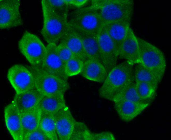
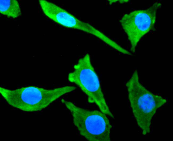
ICC staining of beta Tubulin in SH-SY5Y cells (green). Formalin fixed cells were permeabilized with 0.1% Triton X-100 in TBS for 10 minutes at room temperature and blocked with 10% negative goat serum for 15 minutes at room temperature. Cells were probed with the primary antibody (orb1499401, 1/50) for 1 hour at room temperature, washed with PBS. Alexa Fluor®488 conjugate-Goat anti-Rabbit IgG was used as the secondary antibody at 1/1000 dilution. The nuclear counter stain is DAPI (blue).
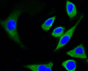
ICC staining of beta Tubulin in SH-SY5Y cells (green). Formalin fixed cells were permeabilized with 0.1% Triton X-100 in TBS for 10 minutes at room temperature and blocked with 10% negative goat serum for 15 minutes at room temperature. Cells were probed with the primary antibody (orb1499401, 1/50) for 1 hour at room temperature, washed with PBS. Alexa Fluor®488 conjugate-Goat anti-Rabbit IgG was used as the secondary antibody at 1/1000 dilution. The nuclear counter stain is DAPI (blue).
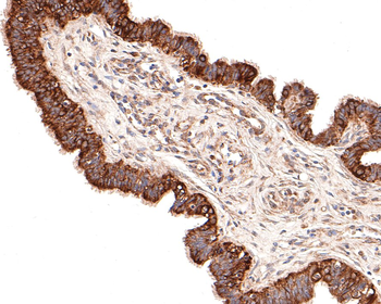
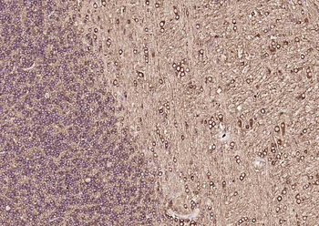
Immunohistochemical analysis of paraffin-embedded human fallopian tube tissue with Rabbit anti-beta Tubulin antibody (orb1499401) at 1/100 dilution. The section was pre-treated using heat mediated antigen retrieval with Tris-EDTA buffer (pH9.0) for 20 minutes. The tissues were blocked in 1% BSA for 20 minutes at room temperature, washed with ddH2O and PBS, and then probed with the primary antibody (orb1499401) at 1/100 dilution for 1 hour at room temperature. The detection was performed using an HRP conjugated compact polymer system. DAB was used as the chromogen. Tissues were counterstained with hematoxylin and mounted with DPX.
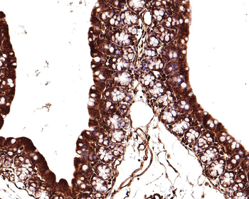
Immunohistochemical analysis of paraffin-embedded mouse large intestine tissue with Rabbit anti-beta Tubulin antibody (orb1499401) at 1/400 dilution. The section was pre-treated using heat mediated antigen retrieval with Tris-EDTA buffer (pH9.0) for 20 minutes. The tissues were blocked in 1% BSA for 20 minutes at room temperature, washed with ddH2O and PBS, and then probed with the primary antibody (orb1499401) at 1/400 dilution for 1 hour at room temperature. The detection was performed using an HRP conjugated compact polymer system. DAB was used as the chromogen. Tissues were counterstained with hematoxylin and mounted with DPX.
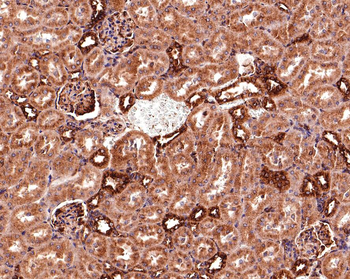
Immunohistochemical analysis of paraffin-embedded rat kidney tissue with Rabbit anti-beta Tubulin antibody (orb1499401) at 1/400 dilution. The section was pre-treated using heat mediated antigen retrieval with Tris-EDTA buffer (pH9.0) for 20 minutes. The tissues were blocked in 1% BSA for 20 minutes at room temperature, washed with ddH2O and PBS, and then probed with the primary antibody (orb1499401) at 1/400 dilution for 1 hour at room temperature. The detection was performed using an HRP conjugated compact polymer system. DAB was used as the chromogen. Tissues were counterstained with hematoxylin and mounted with DPX.
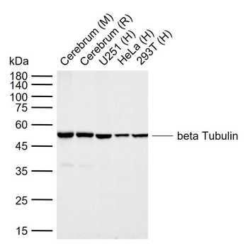
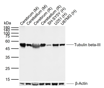
Sample: Lane 1: Mouse Cerebrum tissue lysates, Lane 2: Rat Cerebrum tissue lysates, Lane 3: Human U251 cell lysates, Lane 4: Human HeLa cell lysates, Lane 5: Human 293T cell lysates, Primary: Anti-beta Tubulin (orb1499401) at 1/20000 dilution, Secondary: IRDye800CW Goat Anti-Rabbit IgG at 1/20000 dilution, Predicted band size: 50 kDa, Observed band size: 50 kDa.
Quick Database Links
Gene Symbol
TUBB
UniProt
UniProt Details
− No UniProt data available
Documents Download
Datasheet
Product Information
Request a Document
Protocol Information
WB
Western Blot (IB, immunoblot)
IHC-P
Immunohistochemistry Paraffin
IHC-Fr
Immunohistochemistry Frozen
IF
Immunofluorescence
Beta Tubulin Recombinant Rabbit Monoclonal Antibody (orb1499401)

- 0.0
Based on 0 reviews
Participating in our Biorbyt product reviews program enables you to support fellow scientists by sharing your firsthand experience with our products.
Login to Submit a ReviewAvailable Sizes
Select a size below
Free Secondary Antibody (20 ul)0/0
Please add an antibody product to your cart first.