You have no items in your shopping cart.
Description
Images & Validation
−Item 1 of 3
| Tested Applications | ELISA, IHC, WB |
|---|---|
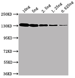
| Dilution Range | ELISA: 1:10,000, IHC: 1:1,500, WB: 1:5,000 - 1:10,000 |
| Application Notes |
Key Properties
−| Antibody Type | Primary Antibody |
|---|---|
| Host | Rabbit |
| Clonality | Polyclonal |
| Isotype | IgG |
| Immunogen | Full length native Beta Galactosidase isolated from E.coli |
| Target | lacZ |
| Purity | Beta-Galactosidase Antibody is an IgG fraction antibody purified from monospecific antiserum by a multi-step process which includes delipidation, salt fractionation and ion exchange chromatography followed by extensive dialysis against the buffer stated above. Assay by immunoelectrophoresis resulted in a single precipitin arc against anti-Rabbit Serum as well as purified and partially purified Beta Galactosidase [E.coli]. Cross reactivity against Beta Galactosidase from other tissues and species may occur but have not been specifically determined. Very low background staining has been reported in various assays. |
| Conjugation | Unconjugated |
Storage & Handling
−| Storage | Store vial at -20° C or below prior to opening. This vial contains a relatively low volume of reagent (25 µL). To minimize loss of volume dilute 1:10 by adding 225 µL of the buffer stated above directly to the vial. Recap, mix thoroughly and briefly centrifuge to collect the volume at the bottom of the vial. Use this intermediate dilution when calculating final dilutions as recommended below. Store the vial at -20°C or below after dilution. Avoid cycles of freezing and thawing. |
|---|---|
| Form/Appearance | Liquid (sterile filtered) |
| Buffer/Preservatives | Preservative: 0.01% (w/v) Sodium Azide. Stabilizer: None; Buffer: 0.02 M Potassium Phosphate, 0.15 M Sodium Chloride, pH 7.2 |
| Concentration | 1.0mg/mL |
| Expiration Date | 12 months from date of receipt. |
| Dry Ice Shipping | Please note: This product requires shipment on dry ice. A dry ice surcharge will apply. |
| Disclaimer | For research use only |
Alternative Names
−rabbit anti-Beta Galactosidase Antibody, rabbit anti-beta gal antibody, ß-Gal, Anti-ß-Gal Antibody
Similar Products
−- Item 1 of 4
- Item 1 of 3
- Item 1 of 1
LASP1 Rabbit Polyclonal Antibody [orb184836]
IF, IHC-Fr, IHC-P
Bovine, Gallus, Mouse, Rabbit, Rat, Sheep
Human
Rabbit
Polyclonal
Unconjugated
50 μl, 100 μl, 200 μl - Item 1 of 2
- Item 1 of 2


Quality Guarantee
Explore bioreagents carefree to elevate your research. All our products are rigorously tested for performance. If a product does not perform as described on its datasheet, our scientific support team will provide expert troubleshooting, a prompt replacement, or a refund. For full details, please see our Terms & Conditions and Buying Guide. Contact us at [email protected].

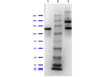
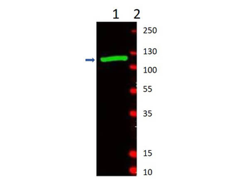
Western Blot of Rabbit Anti-Beta-Galactosidase Antibody. Lane 1: Beta-Galactosidase Reduced [0.1 µg]. Lane 2: Opal Prestained Molecular Weight Marker. Lane 3: Beta-Galactosidase Non-Reduced [0.1 µg]. Primary Antibody: Anti-Beta-Galactosidase at 1:1000 overnight at 2-8°C. Secondary Antibody: Goat Anti-Rabbit IgG HRP (p/n orb347654) 1:70000 for 30 min at RT. Expected MW: ~117kDa.

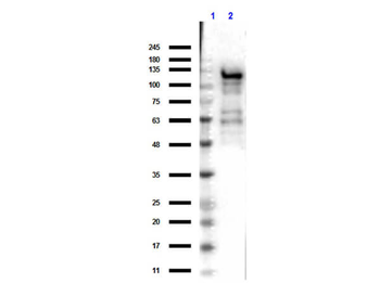
Western Blot of Rabbit Anti-Beta-Galactosidase Antibody. Lane 1: partially purified preparation b-Galactosidase [1.0 µg]. Lane 2: Molecular Weight Marker. Primary Antibody: Anti-Beta-Galactosidase at 1:1000 overnight at 2-8°C. Secondary Antibody: Goat Anti-Rabbit IgG IRDye®800 1:10000 for 45min at RT. Observed MW: ~117kDa.

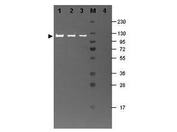
Western blotting using Biorbyt's Fluorescein conjugated anti-b-Galactosidase antibody shows a band at ~117 kDa (lanes 1 - 3) corresponding to 60 ng, 30 ng and 15 ng, respectively of b-Gal present in partially purified preparations (arrowhead). Lane 4 shows no cross reactivity with proteins present in a non-specific control E.coli lysate. Proteins were resolved on a 4-20% Tris-Glycine gel by SDS-PAGE and transferred to nitrocellulose and blocking using Blocking Buffer for Fluorescent Western Blotting (p/n orb348637). The membrane was probed with fluorescein conjugated anti-b-Galactosidase (p/n orb344848) diluted to 1:10000. Reaction occurred for 2 hours at room temperature. Molecular weight estimation was made by comparison to a prestained MW marker in lane M.
Quick Database Links
UniProt Details
− No UniProt data available
NCBI Reference Sequences
−Associated Accession Numbers
Curated reference sequences for the gene transcript and protein product| Protein | NP_414878.1 |
|---|
Documents Download
Datasheet
Product Information
Request a Document
Protocol Information
WB
Western Blot (IB, immunoblot)
IHC
Immunohistochemistry
ELISA
Enzyme-linked Immunosorbent Assay (EIA)
lacZ Antibody (orb344789)

- 0.0
Based on 0 reviews
Participating in our Biorbyt product reviews program enables you to support fellow scientists by sharing your firsthand experience with our products.
Login to Submit a ReviewAvailable Sizes
Select a size below
Free Secondary Antibody (20 ul)0/0
Please add an antibody product to your cart first.