You have no items in your shopping cart.
Description
Research Area
Cancer Biology, Cell Biology, Epigenetics & Chromatin, Signal Transduction
Images & Validation
−Item 1 of 3
| Tested Applications | ICC, IF, WB |
|---|---|
| Dilution Range | WB (1:1000); ICC/IF (1:100) |
| Reactivity | Human, Rat |
| Application Notes |
Key Properties
−| Host | Rabbit |
|---|---|
| Clonality | Polyclonal |
| Immunogen | Synthetic peptide from the mid-protein of human Beclin 1 |
| Target | Beclin 1 |
| Molecular Weight | 30kDa |
| Purification | Peptide Affinity Purified |
| Conjugation | FITC |
Storage & Handling
−| Storage | Conjugated antibodies should be stored according to the product label |
|---|---|
| Buffer/Preservatives | 640.91mM DMSO, 136.36 mM Ethanolamine, 126.89 mM chlorides, 9.09mM phosphates, 9.09mM NaHCO3 |
| Concentration | 1 mg/ml |
| Expiration Date | 12 months from date of receipt. |
| Disclaimer | For research use only |
Alternative Names
−Beclin 1, BECN1, BECN1_HUMAN, APG6, BCL-2 interacting protein beclin, Beclin 1 autophagy related, GT197, VPS30
Similar Products
−- Item 1 of 4
- Item 1 of 1
Beclin 1 Rabbit Polyclonal Antibody (FITC) [orb15177]
IF
Bovine, Porcine
Human, Mouse, Porcine, Rat
Rabbit
Polyclonal
FITC
100 μl - Item 1 of 3

Beclin 1 Rabbit Polyclonal Antibody (FITC) [orb399667]
ICC, IF
Human, Mouse, Rat
Rabbit
Polyclonal
FITC
100 μgAMBRA1 Rabbit Polyclonal Antibody (FITC) [orb7320]
IF
Canine, Equine, Gallus, Human, Mouse, Rabbit
Rat
Rabbit
Polyclonal
FITC
100 μl
Quality Guarantee
Explore bioreagents carefree to elevate your research. All our products are rigorously tested for performance. If a product does not perform as described on its datasheet, our scientific support team will provide expert troubleshooting, a prompt replacement, or a refund. For full details, please see our Terms & Conditions and Buying Guide. Contact us at [email protected].
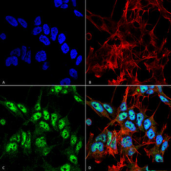
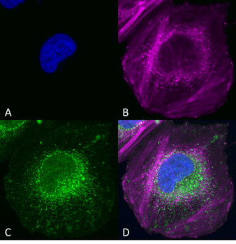
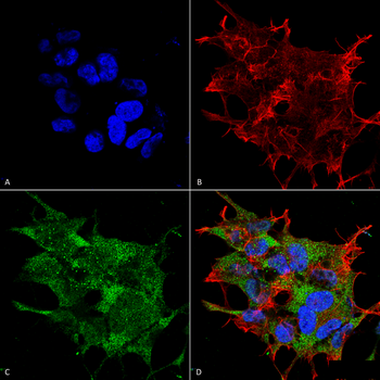
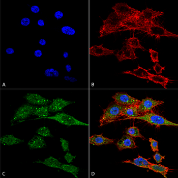
Immunocytochemistry/Immunofluorescence analysis using Rabbit Anti-Beclin 1 Polyclonal Antibody. Tissue: Neuroblastoma cell line (SK-N-BE). Species: Human. Fixation: 4% Formaldehyde for 15 min at RT. Primary Antibody: Rabbit Anti-Beclin 1 Polyclonal Antibody at 1:100 for 60 min at RT. Secondary Antibody: Goat Anti-Rabbit ATTO 488 at 1:100 for 60 min at RT. Counterstain: Phalloidin Texas Red F-Actin stain; DAPI (blue) nuclear stain at 1:1000, 1:5000 for 60min RT, 5min RT. Localization: Nucleus. Magnification: 60X. (A) DAPI (blue) nuclear stain (B) Phalloidin Texas Red F-Actin stain (C) Beclin 1 Antibody (D) Composite.
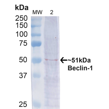
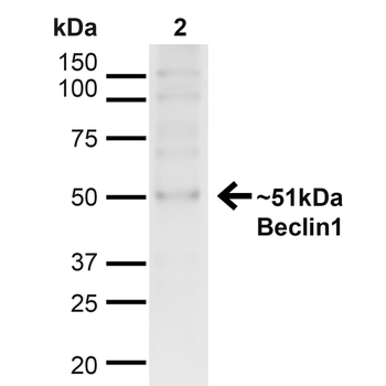
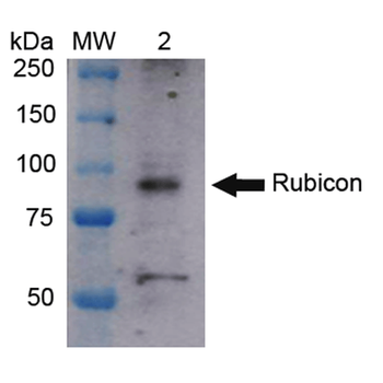
Western blot analysis of Human Cervical cancer cell line (HeLa) lysate showing detection of ~51kDa Beclin 1 protein using Rabbit Anti-Beclin 1 Polyclonal Antibody. Lane 1: MW Ladder. Lane 2: Human HeLa (20 μg). Load: 20 μg. Block: 5% milk + TBST for 1 hour at RT. Primary Antibody: Rabbit Anti-Beclin 1 Polyclonal Antibody at 1:1000 for 1 hour at RT. Secondary Antibody: Goat Anti-Rabbit: HRP at 1:2000 for 1 hour at RT. Color Development: TMB solution for 12 min at RT. Predicted/Observed Size: ~51kDa.
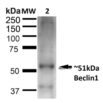
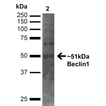
Western blot analysis of Rat Liver showing detection of ~51kDa Beclin 1 protein using Rabbit Anti-Beclin 1 Polyclonal Antibody. Lane 1: MW Ladder. Lane 2: Rat Liver (20 μg). Load: 20 μg. Block: 5% milk + TBST for 1 hour at RT. Primary Antibody: Rabbit Anti-Beclin 1 Polyclonal Antibody at 1:1000 for 1 hour at RT. Secondary Antibody: Goat Anti-Rabbit: HRP at 1:2000 for 1 hour at RT. Color Development: TMB solution for 12 min at RT. Predicted/Observed Size: ~51kDa.
Quick Database Links
UniProt Details
− No UniProt data available
NCBI Gene Details
− No NCBI Gene data available
NCBI Reference Sequences
−Associated Accession Numbers
Curated reference sequences for the gene transcript and protein product| Protein | NP_001300927.1 |
|---|
Documents Download
Datasheet
Product Information
Request a Document
Protocol Information
WB
Western Blot (IB, immunoblot)
IF
Immunofluorescence
ICC
Immunocytochemistry
Beclin 1 Antibody (FITC) (orb377118)

- 0.0
Based on 0 reviews
Participating in our Biorbyt product reviews program enables you to support fellow scientists by sharing your firsthand experience with our products.
Login to Submit a ReviewAvailable Sizes
Select a size below
Choose Conjugation or Carrier Free Version
Free Secondary Antibody (20 ul)0/0
Please add an antibody product to your cart first.