You have no items in your shopping cart.
Description
Research Area
Cell Biology
Images & Validation
−Item 1 of 4
| Tested Applications | IHC-P, WB |
|---|---|
| Dilution Range | WB - 1:1000, IHC-P - 1:25, IF - 1:200 |
| Reactivity | Human, Mouse |
| Predicted Reactivity | Rat |
Key Properties
−| Host | Rabbit |
|---|---|
| Clonality | Polyclonal |
| Isotype | Rabbit IgG |
| Immunogen | This ATG7 antibody is generated from rabbits immunized with a KLH conjugated synthetic peptide between 494-523 amino acids from human ATG7. Antigen Region: 494-523 aa. |
| Target | ATG7 (HGNC:16935) |
| Molecular Weight | 77960 Da |
| Conjugation | Unconjugated |
Storage & Handling
−| Storage | Maintain refrigerated at 2-8°C for up to 2 weeks. For long term storage store at -20°C in small aliquots to prevent freeze-thaw cycles |
|---|---|
| Form/Appearance | Purified polyclonal antibody supplied in PBS with 0.09% (W/V) sodium azide. This antibody is purified through a protein A column, followed by peptide affinity purification. |
| Expiration Date | 12 months from date of receipt. |
| Disclaimer | For research use only |
Alternative Names
−Ubiquitin-like modifier-activating enzyme ATG7, ATG12-activating enzyme E1 ATG7, Autophagy-related protein 7, APG7-like, hAGP7, Ubiquitin-activating enzyme E1-like protein, ATG7, APG7L
Similar Products
−- Item 1 of 8
ATG7 Rabbit Polyclonal Antibody [orb1819451]
ELISA, FC, ICC, IF, IHC, WB
Human
Rabbit
Polyclonal
Unconjugated
100 μg - Item 1 of 6
ATG7 Rabbit Polyclonal Antibody [orb654295]
FC, ICC, IHC, WB
Human, Mouse, Rat
Rabbit
Polyclonal
Unconjugated
100 μg - Item 1 of 5
ATG7 Antibody (C-term) [orb1933432]
IF, IHC-P, WB
Rat
Human, Mouse
Rabbit
Polyclonal
Unconjugated
50 μl, 100 μl - Item 1 of 2
ATG7/APG7 Rabbit Polyclonal Antibody [orb13259]
IF, IHC-Fr, IHC-P, WB
Bovine, Canine, Equine, Gallus, Porcine, Rat
Human, Mouse
Rabbit
Polyclonal
Unconjugated
50 μl, 100 μl, 200 μl - Item 1 of 1
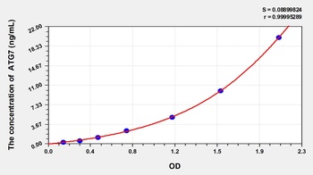
Human Autophagy Related Protein 7 (ATG7) ELISA Kit [orb780922]
Human
0.32-20 ng/mL
0.118 ng/mL
48 T, 96 T
Quality Guarantee
Explore bioreagents carefree to elevate your research. All our products are rigorously tested for performance. If a product does not perform as described on its datasheet, our scientific support team will provide expert troubleshooting, a prompt replacement, or a refund. For full details, please see our Terms & Conditions and Buying Guide. Contact us at [email protected].
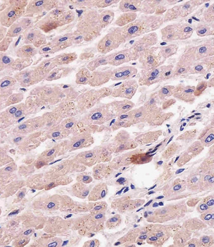
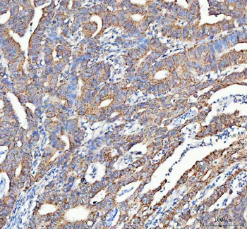
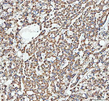
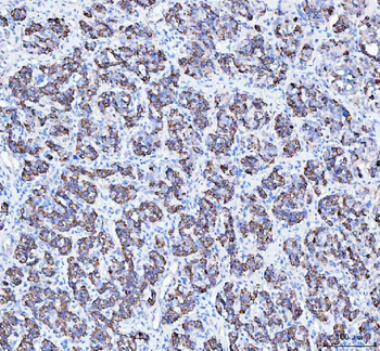
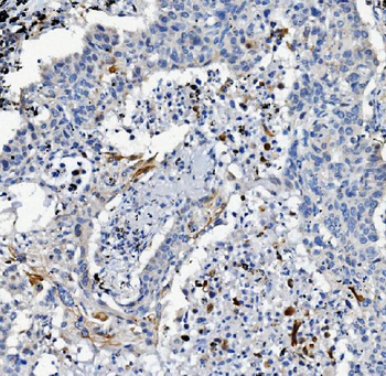
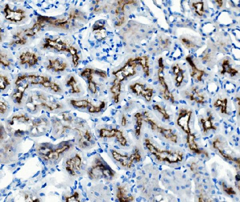
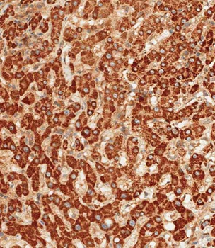
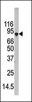
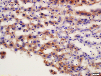
Staining APG7L in human liver tissue sections by Immunohistochemistry (IHC-P - paraformaldehyde-fixed, paraffin-embedded sections). Tissue was fixed with formaldehyde and blocked with 3% BSA for 0.5 hour at room temperature; antigen retrieval was by heat mediation with a citrate buffer (pH6). Samples were incubated with primary antibody (1/25) for 1 hours at 37°C. A undiluted biotinylated goat polyvalent antibody was used as the secondary antibody.
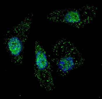
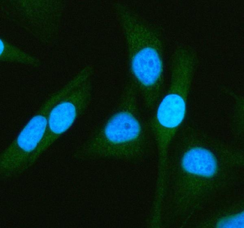
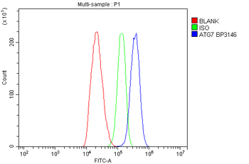
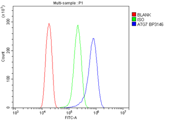
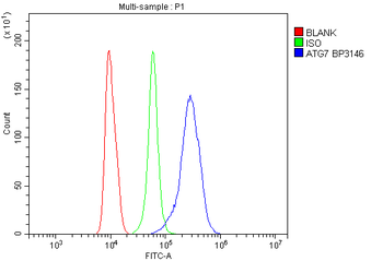
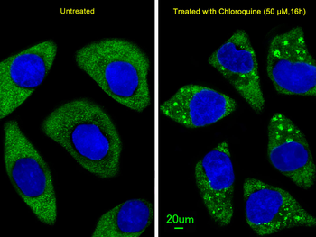
Fluorescent image of U251 cells stained with ATG7 antibody. U251 cells were treated with Chloroquine (50 μM, 16 h), then fixed with 4% PFA (20 min), permeabilized with Triton X-100 (0.2%, 30 min). Cells were then incubated with ATG7 primary antibody (1:200, 2 h at room temperature). For secondary antibody, Alexa Fluor 488 conjugated donkey anti-rabbit antibody (green) was used (1:1000, 1 h). Nuclei were counterstained with Hoechst 33342 (blue) (10 μg/ml, 5 min). ATG7 immunoreactivity is localized to autophagic vacuoles in the cytoplasm of U251 cells.

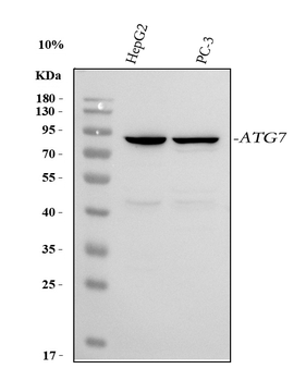
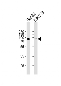
Western blot analysis of anti-hAPG7L-R509 Pab in 293 cell line lysates transiently transfected with the ATG7 gene (2 ug/lane). hAPG7L-R509 (arrow) was detected using the purified Pab.
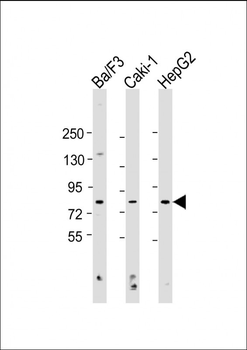
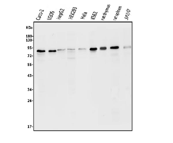
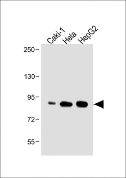
All lanes: Anti-APG7L Antibody (R509) at 1:1000 dilution. Lane 1: Ba/F3 whole cell lysate. Lane 2: Caki-1 whole cell lysate. Lane 3: HepG2 whole cell lysate. Lysates/proteins at 20 µg per lane. Secondary Goat Anti-Rabbit IgG, (H+L), Peroxidase conjugated at 1/10000 dilution. Predicted band size: 78 kDa. Blocking/Dilution buffer: 5% NFDM/TBST.
Quick Database Links
UniProt Details
− No UniProt data available
NCBI Reference Sequences
−Associated Accession Numbers
Curated reference sequences for the gene transcript and protein product| Protein | NP_001129503.2, NP_006386.1, NP_001138384.1 |
|---|
Documents Download
Datasheet
Product Information
Request a Document
Protocol Information
WB
Western Blot (IB, immunoblot)
IHC-P
Immunohistochemistry Paraffin
ATG7 Antibody (orb1933433)

- 0.0
Based on 0 reviews
Participating in our Biorbyt product reviews program enables you to support fellow scientists by sharing your firsthand experience with our products.
Login to Submit a ReviewAvailable Sizes
Select a size below
Choose Conjugation or Carrier Free Version
Free Secondary Antibody (20 ul)0/0
Please add an antibody product to your cart first.