
You have no items in your shopping cart.
Apoptosis Research Hub
Navigate the complete cascade of programmed cell death. Whether you are profiling early membrane asymmetry, tracking caspase execution, or modeling disease with targeted pharmacological inducers.
What defines Early Apoptosis vs. Necrosis, and how do I reliably measure the difference?
Apoptosis is an ATP-dependent, programmed cell death pathway characterized by cell shrinkage, chromatin condensation, and targeted degradation without inducing an inflammatory response. In contrast, classic accidental necrosis is an unprogrammed cell death process resulting in cellular swelling, immediate loss of membrane integrity, and subsequent tissue inflammation, distinguishing it from regulated necrotic modalities like necroptosis.
To differentiate the two, researchers must identify early membrane asymmetry. During early apoptosis, phosphatidylserine (PS) translocates from the inner to the outer leaflet of the plasma membrane. Utilizing Annexin V paired with a membrane-impermeable viability dye (like Propidium Iodide or 7-AAD) allows for precise differentiation using flow cytometry.
Cited Product
Flow Cytometry Kit
Annexin V-FITC Apoptosis Detection Kit
The gold standard for early apoptosis detection. Uses calcium-dependent Annexin V-FITC binding to externalized PS combined with PI.
Target: Phosphatidylserine (PS)
Format: 100 Tests
Application: Flow Cytometry, Microscopy
Essential
Fluorescent Assay
Necrosis vs Apoptosis Kit
Simultaneously monitor apoptotic, necrotic, and healthy cells in a single well using tri-color fluorescence.
Mechanism: PS exposure & Membrane Integrity
Readout: Fluorometric / Plate Reader
Application: High-throughput screening
How do I profile the Intrinsic (Mitochondrial) Apoptosis Pathway in my disease models?
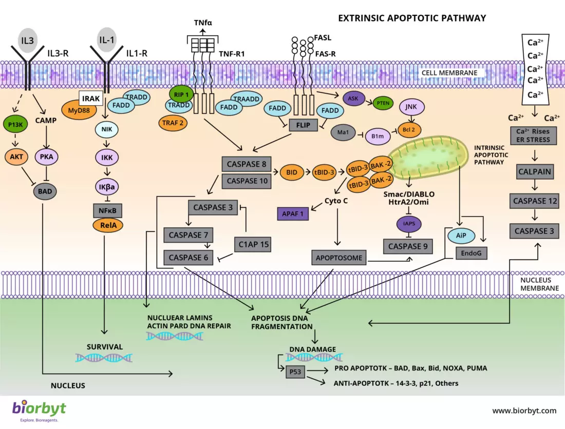
The intrinsic pathway is activated by intracellular stress (e.g., DNA damage, oxidative stress) and is tightly governed by the Bcl-2 family of proteins at the mitochondrial outer membrane. A shift in the ratio of anti-apoptotic factors (Bcl-2, Bcl-xL) to pro-apoptotic factors (Bax, Bak) leads to Mitochondrial Outer Membrane Permeabilization (MOMP) and the release of Cytochrome c.
Additionally, the accumulation of specific bioactive sphingolipids, particularly Ceramides, directly triggers mitochondrial depolarization by forming channels in the outer mitochondrial membrane or synergizing with pro-apoptotic Bcl-2 proteins. Profiling these lipid-protein signaling axes requires highly sensitive quantification tools.
Cited Product
ELISA Kit
Rat BCL-2 ELISA Kit
Quantify anti-apoptotic Bcl-2 levels to monitor intrinsic pathway activation. (Cited in PMC10609711 for neurodegeneration models).
Featured
Competitive ELISA
Ceramide ELISA Kit
Measure ceramide levels, critical second messengers in lipid-induced apoptosis. (Cited in PMC12172293 for hepatic steatosis).
Sensitivity: 18.75 pg/ml
Range: 31.2 - 2000 pg/ml
Application: Lipid Signaling Analysis
How do tumor cells evade cell death, and what regulatory markers should I target?
Cancer cells frequently evade apoptosis by upregulating Inhibitor of Apoptosis Proteins (IAPs) or downregulating pro-apoptotic signaling networks. A primary target is Survivin (BIRC5), a prominent IAP that indirectly suppresses cell death by stabilizing XIAP and neutralizing the pro-apoptotic factors Smac/DIABLO. Survivin is heavily overexpressed in most human tumors but practically absent in normal, terminally differentiated cells.
Furthermore, the loss or epigenetic silencing of SFRP1 (Secreted Frizzled-Related Protein 1) releases canonical Wnt/β-catenin signaling from inhibition, promoting unrestrained cell survival and apoptosis evasion.
Precision Target
Sandwich ELISA
Human Survivin ELISA Kit
Accurately quantify BIRC5/Survivin concentrations to assess anti-apoptotic signaling and tumor resistance.
Sensitivity: < 2 pg/ml
Range: 62.5 - 4000 pg/ml
Target: BIRC5 (IAP Family)
Precision Target
Sandwich ELISA
Human SFRP1 ELISA Kit
Measure soluble SFRP1 levels to investigate Wnt pathway antagonism and its subsequent role in apoptosis induction.
Sensitivity: < 0.05 ng/ml
Sample: Supernatant, Serum, Plasma
Pathway: Wnt / β-catenin
What are the best assays for Late-Stage Execution and how can I reliably induce it?
Once initiator caspases (-8 or -9) activate executioner caspases (-3, -6, -7), the cell is irreversibly committed to death. Late-stage markers include the cleavage of PARP and internucleosomal fragmentation of DNA (yielding ~200bp ladders), which is best detected using TUNEL assays.
To establish a robust positive control in in vitro models, researchers utilize highly pure pharmacological modulators like 5-Fluorouracil (which triggers p53 (WT)-dependent apoptosis via DNA damage, a process tightly regulated by proteins like phospho-MDM2 and specific modifications such as p53 Ser392 phosphorylation) or Streptozocin (a DNA alkylating agent). Evaluating cells with mutant p53 profiles is critical in these assays to verify targeted pathway induction.
Multiplex
Primary Antibodies
Apoptosis Antibody Panel
A curated suite of primary antibodies covering Caspase-3, Caspase-8, Caspase-9, PARP1, and BAX for cohesive Western Blotting.
Reactivity: Human, Mouse, Rat
Application: WB, IHC, ICC
High Purity
Antimetabolite
5-Fluorouracil (5-FU)
Potent thymidylate synthase inhibitor used globally as a reliable apoptosis inducer in oncology models.
Purity: ≥ 99.0% (HPLC)
Pathway: p53 / DNA Damage
Modality Selection Guide
Analytical Instrument | Optimal Apoptosis Targets | Recommended Biorbyt Solutions |
|---|---|---|
Flow Cytometer | Single-cell quantification of membrane asymmetry, sub-G1 DNA content, and active caspases. | |
Microplate Reader | High-throughput quantification of anti-apoptotic proteins, enzymatic activity, and lipids. | Rat BCL-2 ELISA (orb567225), Ceramide ELISA (orb1881455), Survivin ELISA (orb50135). |
Western Blot / Imaging | Detection of protein cleavage events (PARP, Caspases) and in situ DNA fragmentation. | Apoptosis Antibody Panels (orb3130882), TUNEL Apoptosis Detection Kit (Orange Fluorescence). |
Require a Custom Assay Formulation?
If your cell death research requires specialized reagents that aren't available off-the-shelf, Biorbyt’s team is ready to assist. We provide custom polyclonal/monoclonal antibody generation, specific peptide synthesis, and bespoke assay conjugations to ensure your experimental success.
Scientific Bibliography & Key Citations
- Mustafa, M., et al. (2024). Apoptosis: A Comprehensive Overview of Signaling Pathways, Morphological Changes, and Physiological Significance. Cells, 13(22), 1838. doi:10.3390/cells13221838
- Siskind, L. J. (2005). Mitochondrial ceramide channels: an early stage of apoptosis? Journal of Bioenergetics and Biomembranes, 37(3), 143-153.
- Gelebart, P., et al. (2008). Epigenetic silencing of SFRP1 activates the Wnt/β-catenin signaling pathway and contributes to oncogenesis. Blood, 112(10), 4192-4201.
- Abd Elmaaboud, M. A., et al. (2023). Dapagliflozin/Hesperidin Combination Mitigates Lipopolysaccharide-Induced Alzheimer's Disease in Rats. Pharmaceuticals, 16(11), 1530. (Cites Biorbyt Rat Bcl-2 ELISA orb567225) PMC10609711
- Phelps, C. B., et al. (2024). Genetic depletion of adipose-derived Isthmin-1 causes hepatic steatosis. Molecular Metabolism. (Cites Biorbyt Ceramide ELISA orb1881455) PMC12172293
- Taylor, R. C., Cullen, S. P., & Martin, S. J. (2008). Apoptosis: controlled demolition at the cellular level. Nature Reviews Molecular Cell Biology, 9(3), 231-241. doi:10.1038/nrm2312