You have no items in your shopping cart.
Description
Research Area
Infectious Disease & Virology, Microbiology, Signal Transduction
Images & Validation
−Item 1 of 6
| Tested Applications | IF, IHC-P, WB |
|---|---|
| Dilution Range | IF - 1:10-50, WB - 1:2000, IHC-P - 1:10-50, IHC-P-Leica - 1:500 |
| Reactivity | Human, Mouse |
| Predicted Reactivity | Rat |
Key Properties
−| Host | Rabbit |
|---|---|
| Clonality | Polyclonal |
| Isotype | Rabbit IgG |
| Immunogen | This AP1M1 antibody is generated from rabbits immunized with a KLH conjugated synthetic peptide between 199-227 amino acids from the Central region of human AP1M1. Antigen Region: 199-227 aa. |
| Target | AP1M1 |
| Molecular Weight | 48587 Da |
| Conjugation | Unconjugated |
Storage & Handling
−| Storage | Maintain refrigerated at 2-8°C for up to 2 weeks. For long term storage store at -20°C in small aliquots to prevent freeze-thaw cycles |
|---|---|
| Form/Appearance | Purified polyclonal antibody supplied in PBS with 0.09% (W/V) sodium azide. This antibody is purified through a protein A column, followed by peptide affinity purification. |
| Expiration Date | 12 months from date of receipt. |
| Disclaimer | For research use only |
Alternative Names
−AP-1 complex subunit mu-1, AP-mu chain family member mu1A, Adaptor protein complex AP-1 subunit mu-1, Adaptor-related protein complex 1 subunit mu-1, Clathrin assembly protein complex 1 mu-1 medium chain 1, Clathrin coat assembly protein AP47, Clathrin coat-associated protein AP47, Golgi adaptor HA1/AP1 adaptin mu-1 subunit, Mu-adaptin 1, Mu1A-adaptin, AP1M1, CLTNM
Similar Products
−- Item 1 of 3
AP1M1 Antibody (Center) [orb1938252]
FC, IF, WB
Mouse, Rat
Human
Rabbit
Polyclonal
Unconjugated
50 μl, 100 μl
Quality Guarantee
Explore bioreagents carefree to elevate your research. All our products are rigorously tested for performance. If a product does not perform as described on its datasheet, our scientific support team will provide expert troubleshooting, a prompt replacement, or a refund. For full details, please see our Terms & Conditions and Buying Guide. Contact us at [email protected].
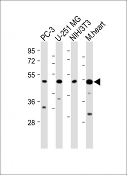
All lanes: Anti-AP1M1 Antibody (Center) at 1:2000 dilution. Lane 1: PC-3 whole cell lysate. Lane 2: U-251 MG whole cell lysate. Lane 3: NIH/3T3 whole cell lysate. Lane 4: Mouse heart lysate.Lysates/proteins at 20 µg per lane. Secondary Goat Anti-Rabbit IgG, (H+L), Peroxidase conjugated at 1/10000 dilution. Predicted band size: 49 kDa. Blocking/Dilution buffer: 5% NFDM/TBST.
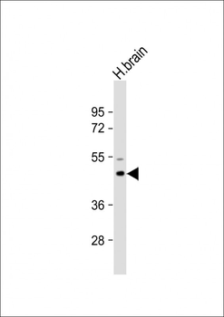
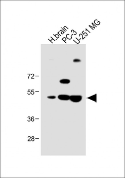
Anti-AP1M1 Antibody (Center) at 1:2000 dilution + Human brain lysate.Lysates/proteins at 20 µg per lane. Secondary Goat Anti-Rabbit IgG, (H+L), Peroxidase conjugated at 1/10000 dilution. Predicted band size: 49 kDa. Blocking/Dilution buffer: 5% NFDM/TBST.
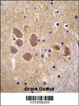
AP1M1 Antibody (Center) immunohistochemistry analysis in formalin fixed and paraffin embedded human brain tissue followed by peroxidase conjugation of the secondary antibody and DAB staining. This data demonstrates the use of AP1M1 Antibody (Center) for immunohistochemistry. Clinical relevance has not been evaluated.
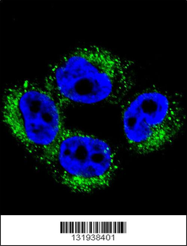
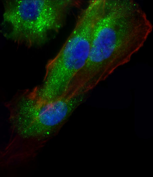
Confocal immunofluorescent analysis of AP1M1 Antibody (Center) with A375 cell followed by Alexa Fluor 488-conjugated goat anti-rabbit lgG (green). DAPI was used to stain the cell nuclear (blue).
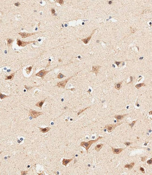
Immunohistochemical analysis of paraffin-embedded human brain tissue performed on the Leica BOND RXm. Samples were incubated with primary antibody (1/500) for 1 hours at room temperature. A undiluted biotinylated CRF Anti-Polyvalent HRP Polymer antibody was used as the secondary Antibody.
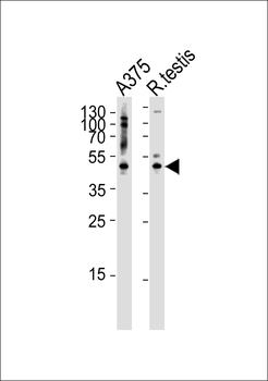
Western blot analysis of lysates from A375 cell line and rat testis tissue lysate (from left to right), using AP1M1 Antibody (Center). diluted at 1:1000 at each lane. A goat anti-rabbit IgG H&L (HRP) at 1:5000 dilution was used as the secondary Antibody. Lysates at 35 ug per lane.
Quick Database Links
UniProt Details
− No UniProt data available
NCBI Reference Sequences
−Associated Accession Numbers
Curated reference sequences for the gene transcript and protein product| Protein | NP_001123996.1, NP_115882.1 |
|---|
Documents Download
Datasheet
Product Information
Request a Document
Protocol Information
WB
Western Blot (IB, immunoblot)
IHC-P
Immunohistochemistry Paraffin
IF
Immunofluorescence
AP1M1 Antibody (Center) (orb1937776)

- 0.0
Based on 0 reviews
Participating in our Biorbyt product reviews program enables you to support fellow scientists by sharing your firsthand experience with our products.
Login to Submit a ReviewAvailable Sizes
Select a size below
Choose Conjugation or Carrier Free Version
Free Secondary Antibody (20 ul)0/0
Please add an antibody product to your cart first.