Queen's Award Received in 2021 ISO 9001 Certified Delivered over 1,000,000 bio-reagents to life science researchers Trusted by Life Science Communities
Cart summary

You have no items in your shopping cart.

SHANK1a Antibody

SKU: orb1821883

Description

Our SHANK1a rabbit polyclonal primary antibody from Biorbyt is produced in-house. It detects human, mouse, and rat SHANK1a and is antigen affinity purified.

Research Area

Neuroscience, Protein Biochemistry, Signal Transduction

Images & Validation

Tested ApplicationsWB
Dilution RangeWB: 1:500-1:1000
ReactivityHuman, Mouse, Rat

Key Properties

Antibody TypePrimary Antibody
HostRabbit
ClonalityPolyclonal
IsotypeIgG
ImmunogenSynthetic peptide corresponding to amino acids from the C-terminal region of human Shank1a protein
TargetSHANK1a
PurificationAntigen Affinity Purified
ConjugationUnconjugated

Storage & Handling

StorageMaintain refrigerated at 2-8°C for up to 2 weeks. For long term storage store at -20°C in small aliquots to prevent freeze-thaw cycles.
Form/AppearanceLiquid
Buffer/PreservativesPBS + 0.02% NaN3
Expiration Date12 months from date of receipt.
DisclaimerFor research use only

Alternative Names

SH3 and multiple ankyrin repeat domains 1, GKAP/SAPAP interacting protein, Somatostatin receptor interacting protein, SSTR interacting protein, SSTRIP, SPANK1

Similar Products

  • SHANK1 Antibody (APC) [orb148619]

    AM,  IHC,  WB

    Human, Mouse, Rat

    Mouse

    Monoclonal

    APC

    100 μg
  • SHANK1 Antibody (Biotin) [orb148620]

    AM,  IHC,  WB

    Human, Mouse, Rat

    Mouse

    Monoclonal

    Biotin

    100 μg
  • SHANK1 Antibody (HRP) [orb148622]

    AM,  IHC,  WB

    Human, Mouse, Rat

    Mouse

    Monoclonal

    HRP

    100 μg
  • SHANK1 Antibody (PerCP) [orb148624]

    AM,  IHC,  WB

    Human, Mouse, Rat

    Mouse

    Monoclonal

    PerCP

    100 μg
  • SHANK1 Antibody (RPE) [orb148625]

    AM,  IHC,  WB

    Human, Mouse, Rat

    Mouse

    Monoclonal

    RPE

    100 μg
Quality Guarantee

Quality Guarantee

Explore bioreagents carefree to elevate your research. All our products are rigorously tested for performance. If a product does not perform as described on its datasheet, our scientific support team will provide expert troubleshooting, a prompt replacement, or a refund. For full details, please see our Terms & Conditions and Buying Guide. Contact us at [email protected].

SHANK1a Antibody

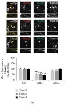
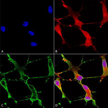
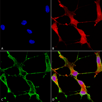
Immunocytochemistry of hippocampal neurons DIV 14 grown under control, ASD1, and ASD2 conditions. The fluorescence intensity of Shank positive puncta was measured using antibodies specific for Shank1, Shank2, and Shank3. Exemplary images (upper panel) and quantification of average puncta signal intensity of 10 cells per condition (lower panel). Merged images show additional DAPI staining of the nucleus.

SHANK1a Antibody

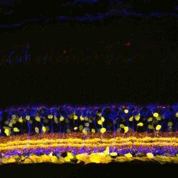
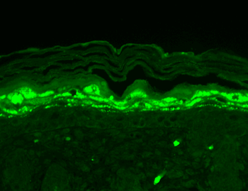
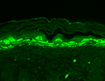
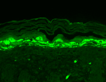
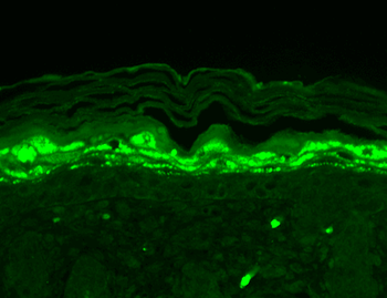
Immunohistochemistry of mouse retinal sections showing specific staining of SHANK1.

SHANK1a Antibody

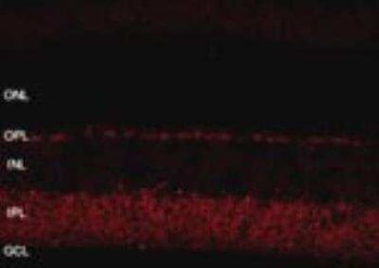
Immunohistochemistry of retinal tissue showing specific staining of Shank at 1:800 dilution. Images are 2048 X 2048, 12 bit, collected with a 40X water objective, 10 stacks 0.55 um per stack.

SHANK1a Antibody

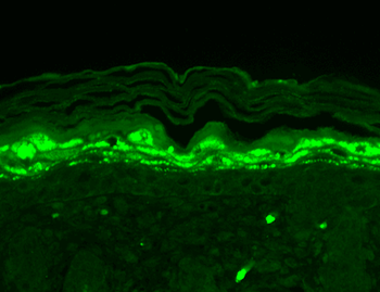
Immunohistochemistry of retinal tissue showing specific staining of Shank at 1:800 dilution. Images are 2048 X 2048, 12 bit, collected with a 40X water objective, 10 stacks 0.55 um per stack.

SHANK1a Antibody

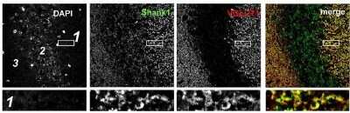
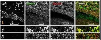
Immunohistochemistry showing colocalization of Shank puncta with VGLUT1 puncta in the intragranular mossy fibers and the stratum moleculare of the dentate gyrus. Confocal immunofluorescence stainings of coronal sections from wild-type mice probed with the Shank1 (white; green in merge) and VGLUT1 (white; red in merge) antibodies. The upper rows (large squares) show the enlarged region (3 = SM; 2 = SG; 1 = PL; scale bar = 100 um), the bottom rows (small rectangles) show further enlargements indicated in up.

SHANK1a Antibody

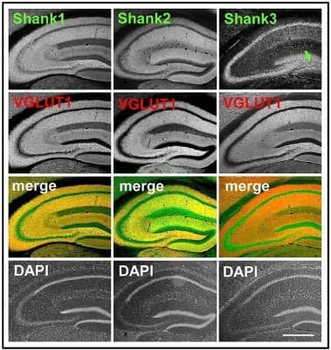
Immunohistochemistry showing synaptic distribution of Shank1/2 and somato-synaptic distribution of Shank3 in the mouse hippocampus, codistribution with VGLUT1, no codistribution with VGLUT2. 5x magnification of hippocampus. Immunofluorescence staining of coronal sections from wild-type mice probed with Shank1-3 (white; green in merge) and VGLUT1 (white; red in merge) antibodies; green arrow points toward the intragranular mossy fibers where there is a prominent synaptic stain of Shank3; scale bar =300 um.

SHANK1a Antibody

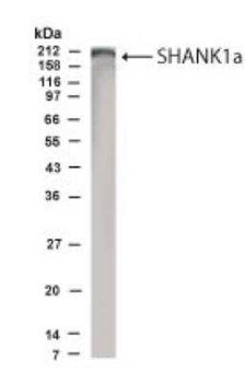
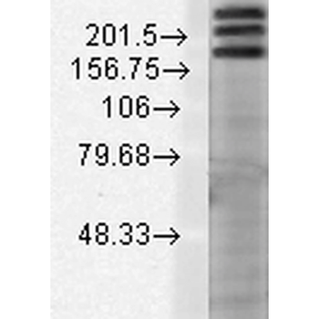
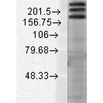
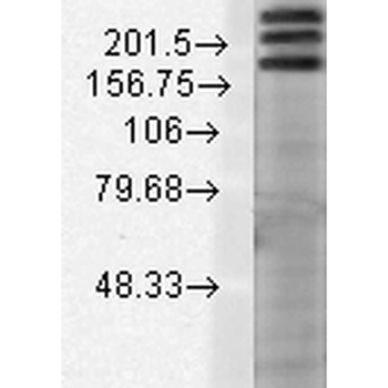
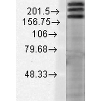
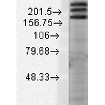
Western Blot of rat brain lysate showing specific immunolabeling of SHANK1 at 1:500 dilution.

UniProt Details

No UniProt data available

Documents Download

Datasheet
Product Information
Download

Request a Document

Protocol Information

WB
Western Blot (IB, immunoblot)
View Protocol

SHANK1a Antibody (orb1821883)

  • Star
  • Star
  • Star
  • Star
  • Star
  • 0.0
Based on 0 reviews

Participating in our Biorbyt product reviews program enables you to support fellow scientists by sharing your firsthand experience with our products.

Login to Submit a Review

No reviews yet

Available Sizes

Select a size below

100 μl
$ 630.00
Choose Conjugation or Carrier Free Version
Free Secondary Antibody (20 ul)0/0

Please add an antibody product to your cart first.

DispatchUsually dispatched within 5-10 working days
Bulk Enquiry