You have no items in your shopping cart.
Featured
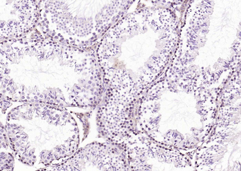
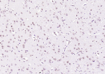
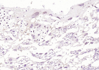
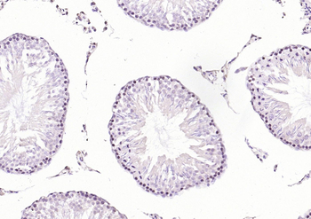
Description
Research Area
Signal Transduction
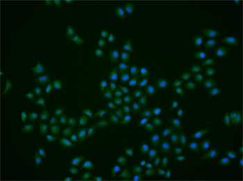
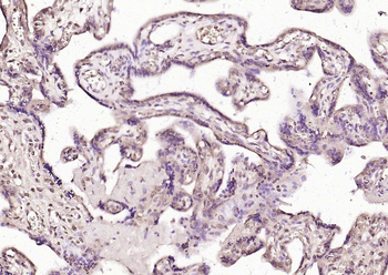
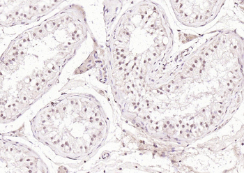
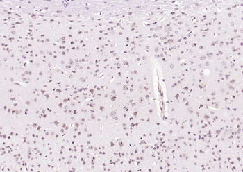
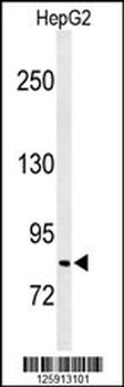
Images & Validation
−
Item 1 of 3
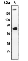
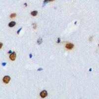
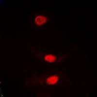
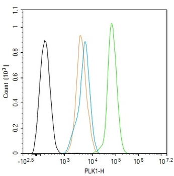
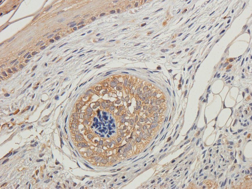
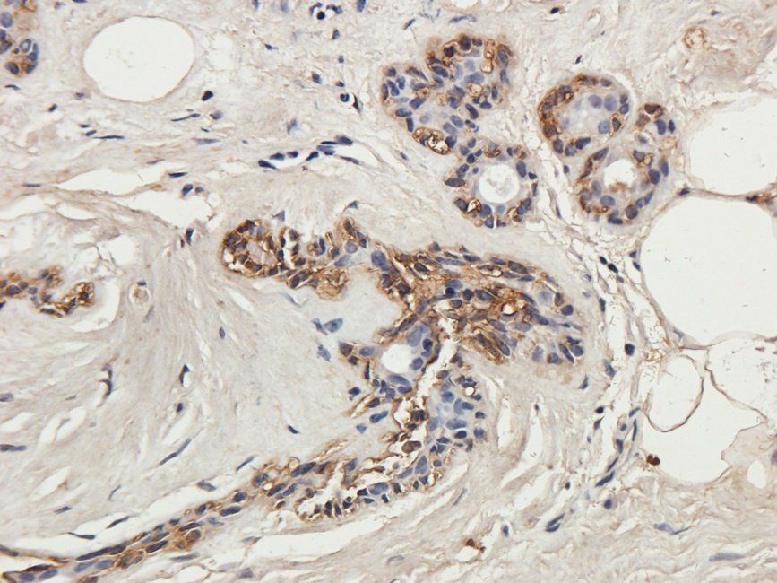
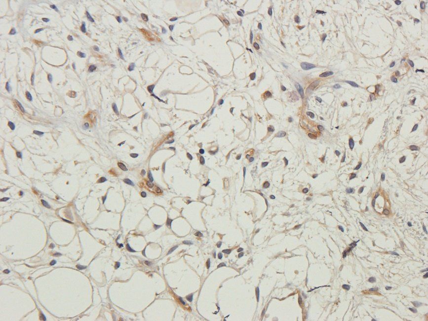
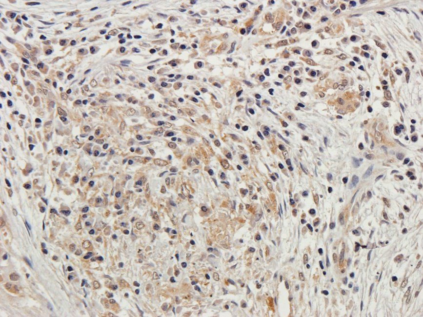
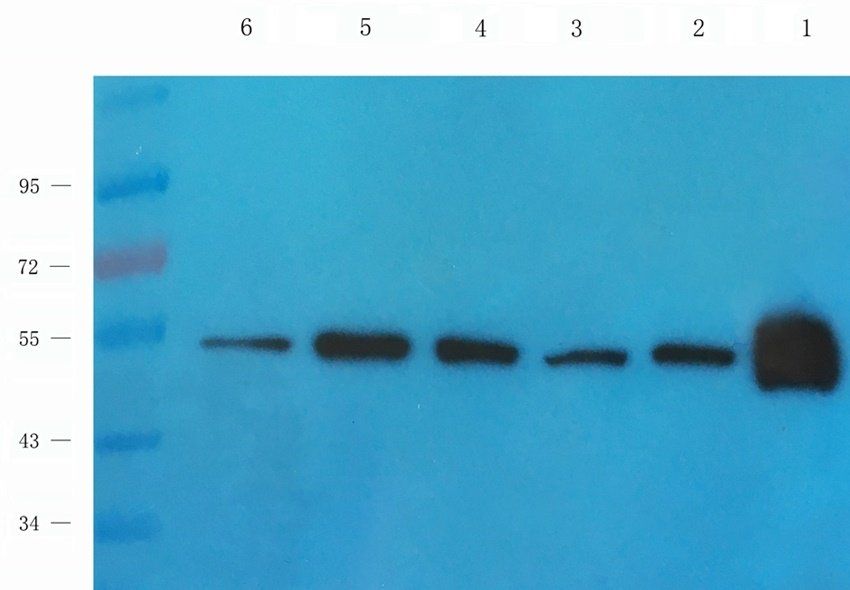
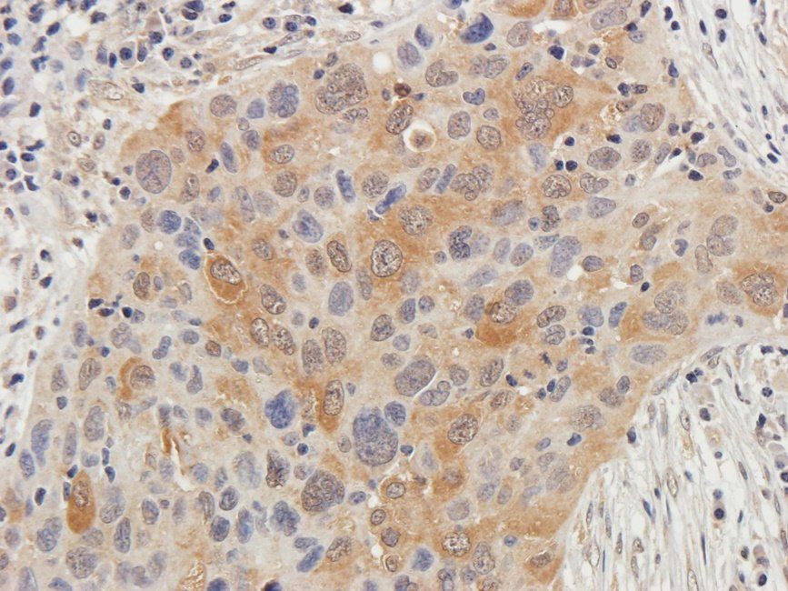
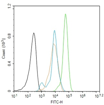
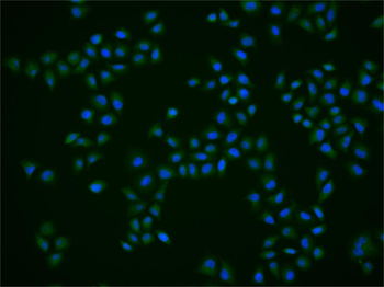
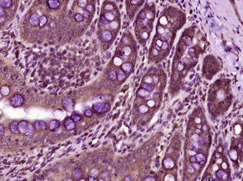
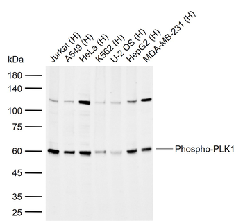
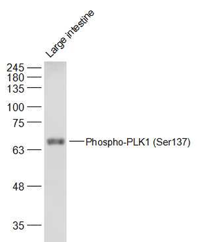
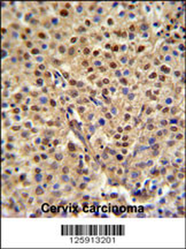
| Tested Applications | IF, IHC, WB |
|---|---|
| Dilution Range | WB: 1-500:1000, IHC-P: 1-100:200, IF/ICC: 1-100:500 |
| Reactivity | Human, Mouse, Rat |
Key Properties
−| Antibody Type | Primary Antibody |
|---|---|
| Host | Rabbit |
| Clonality | Polyclonal |
| Immunogen | KLH-conjugated synthetic peptide encompassing a sequence within the center region of human PLK1. The exact sequence is proprietary. |
| Target | PLK1 |
| Purification | The antibody was purified by immunogen affinity chromatography. |
| Conjugation | Unconjugated |
Storage & Handling
−| Storage | Maintain refrigerated at 2-8°C for up to 2 weeks. For long term storage store at -20°C in small aliquots to prevent freeze-thaw cycles. |
|---|---|
| Form/Appearance | Liquid |
| Buffer/Preservatives | 0.42% Potassium phosphate, 0.87% Sodium chloride, pH 7.3, 30% glycerol, and 0.01% sodium azide. |
| Expiration Date | 12 months from date of receipt. |
| Disclaimer | For research use only |
Alternative Names
−PLK; Serine/threonine-protein kinase PLK1; Polo-like kinase 1; PLK-1; Serine/threonine-protein kinase 13; STPK13
Similar Products
−- Item 1 of 10
PLK1 Rabbit Polyclonal Antibody [orb6761]
FC, IF, IHC-Fr, IHC-P
Canine, Porcine, Rabbit
Human, Mouse, Rat
Rabbit
Polyclonal
Unconjugated
50 μl, 100 μl, 200 μl - Item 1 of 6
- Item 1 of 5
Phospho-PLK1 (Ser137) Rabbit Polyclonal Antibody [orb6763]
FC, IF, IHC-Fr, IHC-P, WB
Bovine, Canine, Gallus, Mouse, Porcine, Rabbit
Human, Rat
Rabbit
Polyclonal
Unconjugated
100 μl, 200 μl, 50 μl - Item 1 of 3
Plk rabbit pAb Antibody [orb769495]
ELISA, IF, IHC, WB
Human, Mouse, Rat
Polyclonal
Unconjugated
50 μl, 100 μl - Item 1 of 3


Quality Guarantee
Explore bioreagents carefree to elevate your research. All our products are rigorously tested for performance. If a product does not perform as described on its datasheet, our scientific support team will provide expert troubleshooting, a prompt replacement, or a refund. For full details, please see our Terms & Conditions and Buying Guide. Contact us at [email protected].
Quick Database Links
UniProt Details
− No UniProt data available
NCBI Gene Details
− No NCBI Gene data available
Protocol Information
WB
Western Blot (IB, immunoblot)
IHC
Immunohistochemistry
IF
Immunofluorescence
Guan, Jianmin et al. Long non‑coding RNA ZEB2‑AS1 affects cell proliferation and apoptosis via the miR‑122‑5p/PLK1 axis in acute myeloid leukemia Int. J. Mol. Med., (2020)
Available Sizes
Select a size below
Choose Conjugation or Carrier Free Version
Free Secondary Antibody (20 ul)0/0
Please add an antibody product to your cart first.