You have no items in your shopping cart.
Featured
Description
Research Area
Apoptotic, Cancer, Epigenetics, Neuroscience, Signaling Pathways
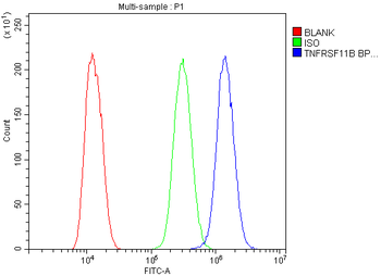
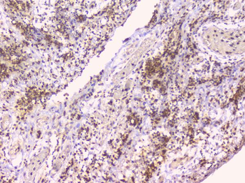
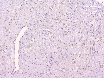
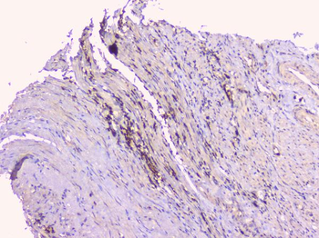
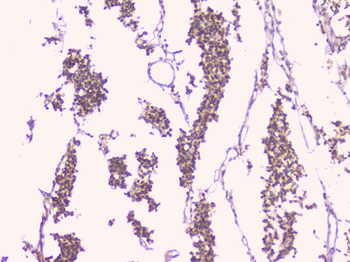
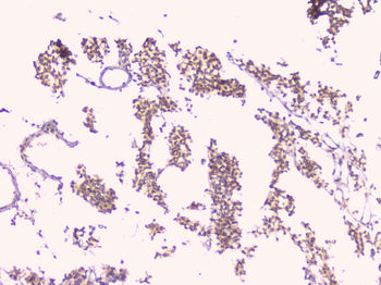
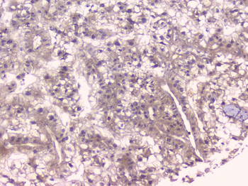
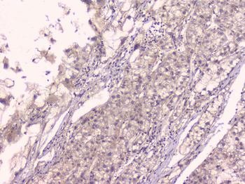
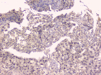
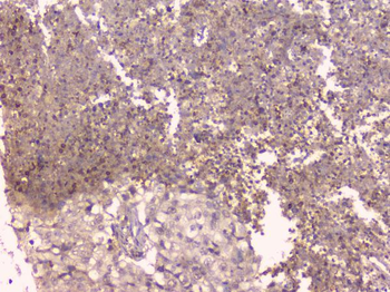
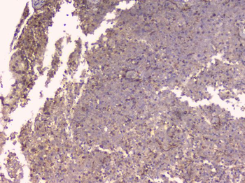
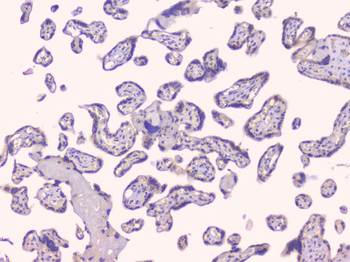
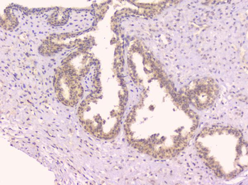
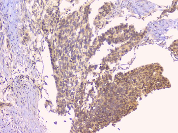
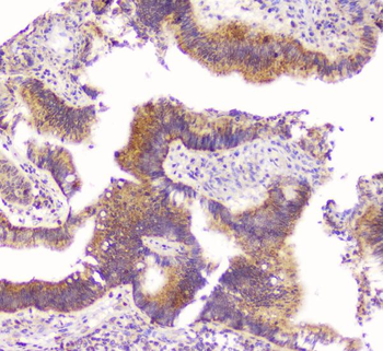
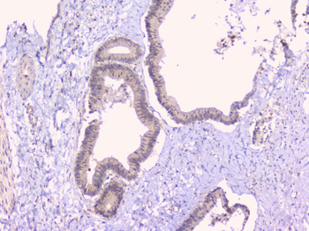
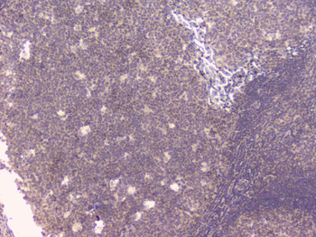
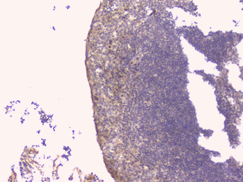
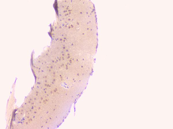
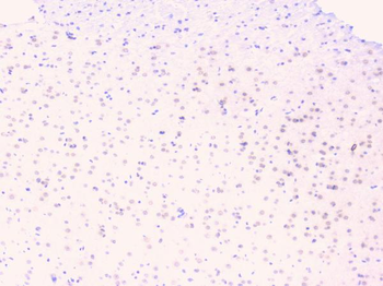
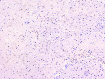
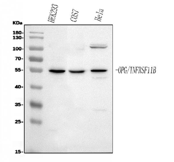
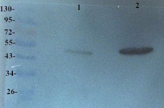
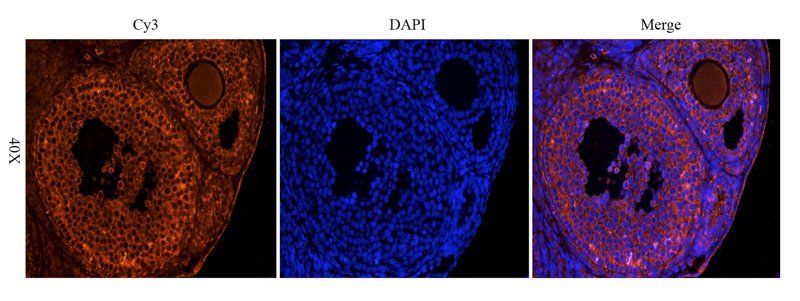
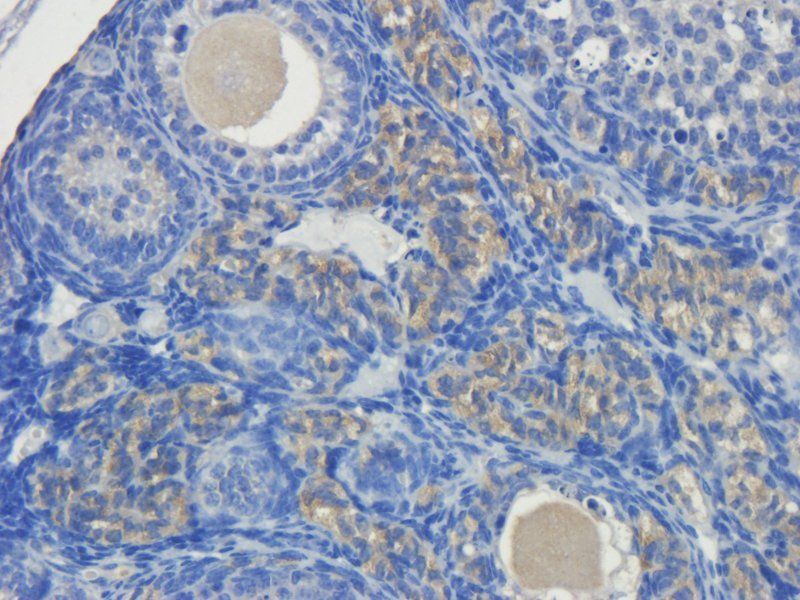
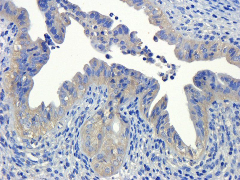
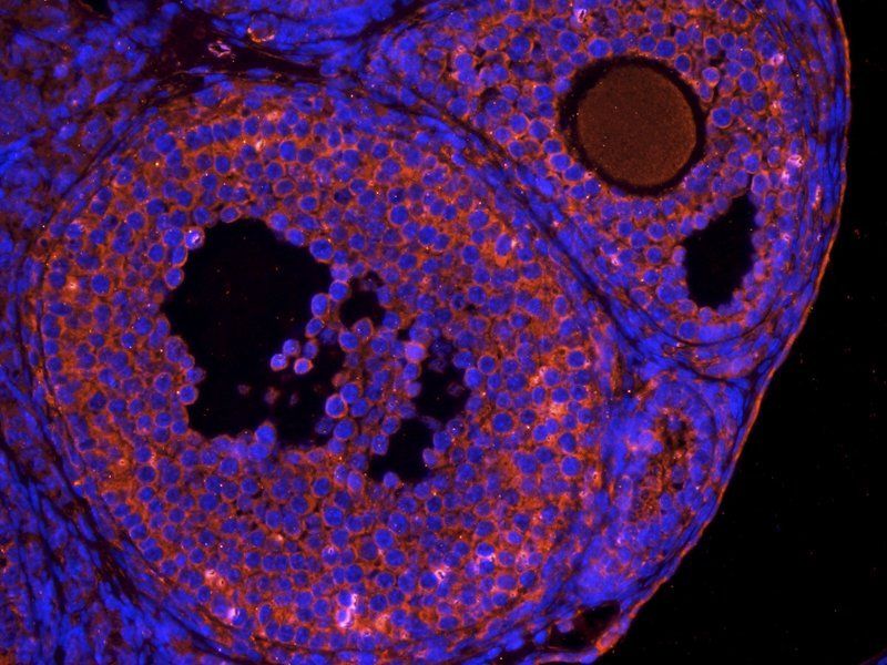
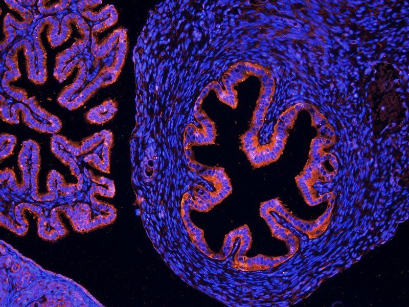
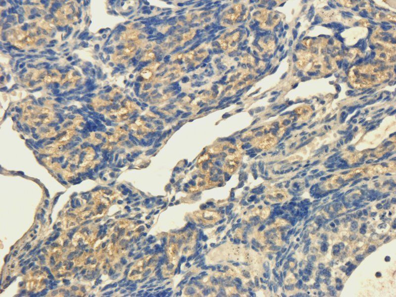
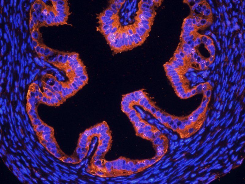
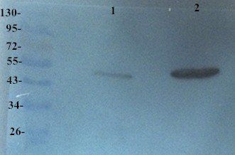
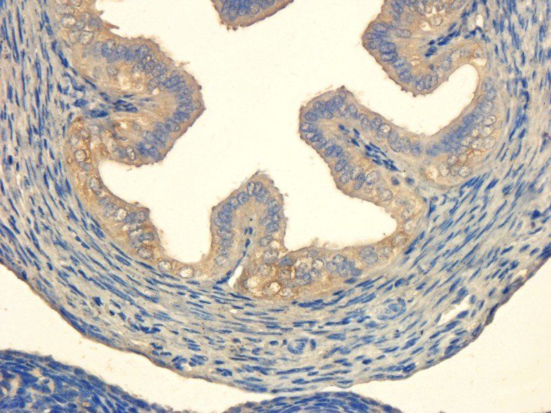
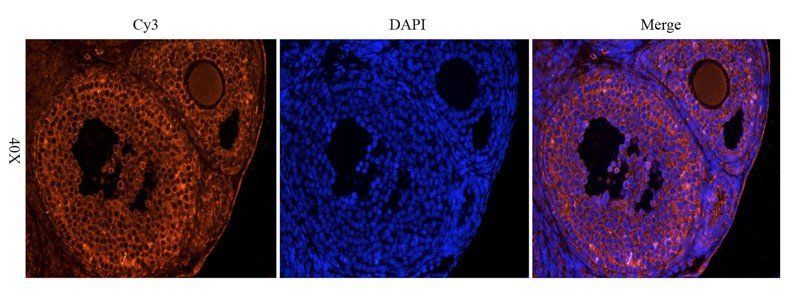
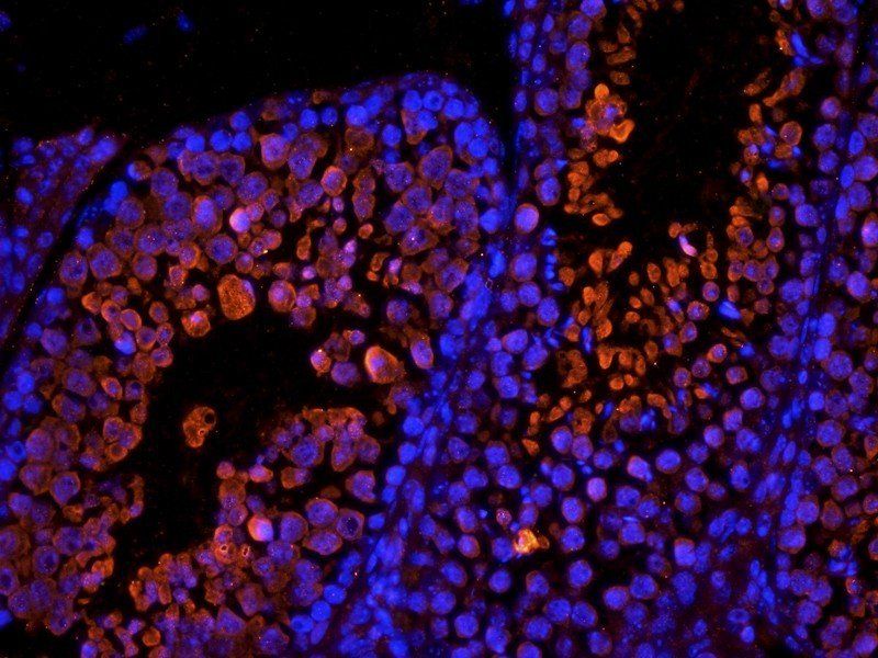
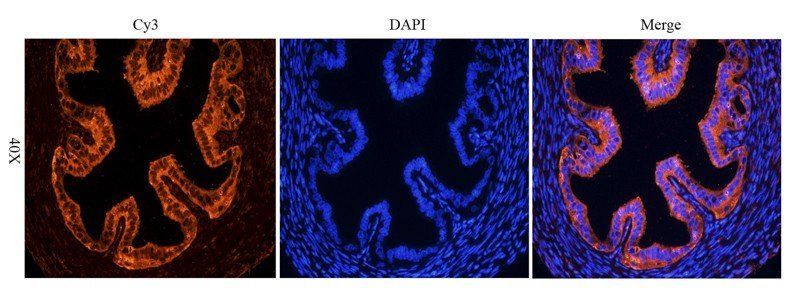
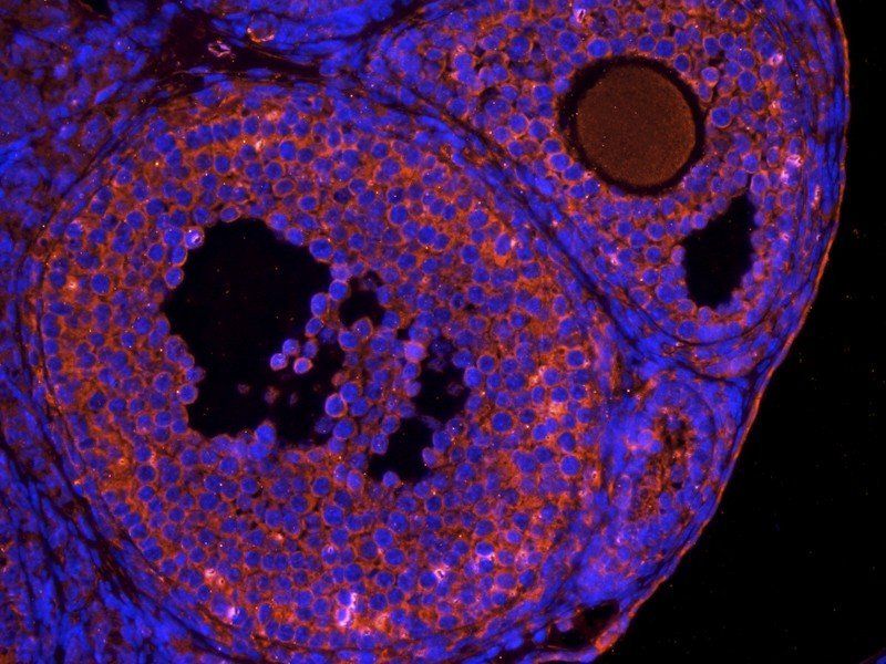
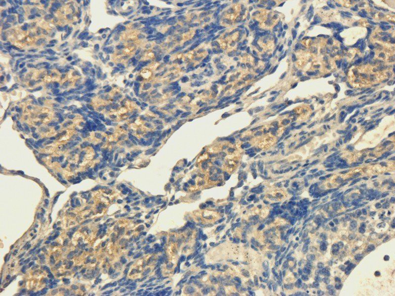
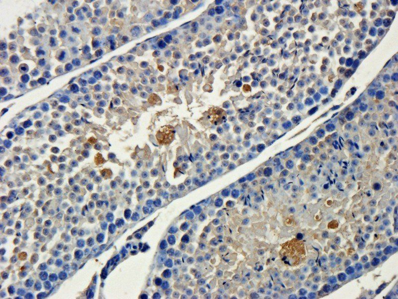
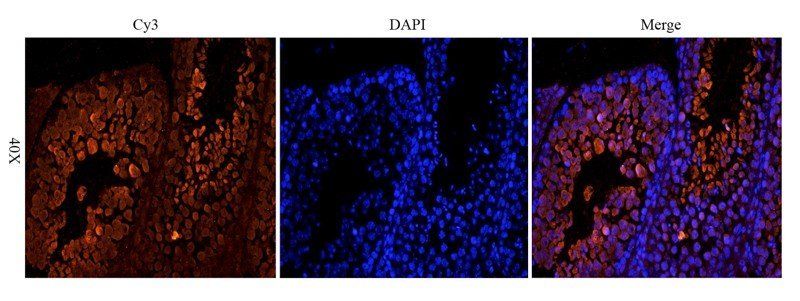
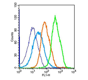
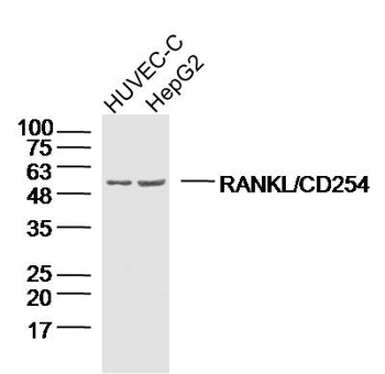
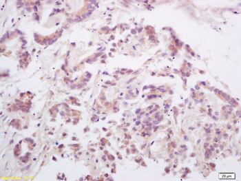
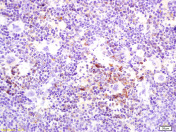
Images & Validation
−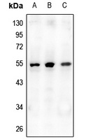
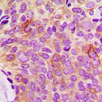
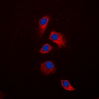
Item 1 of 3
| Tested Applications | IF, IHC, WB |
|---|---|
| Dilution Range | WB: 1:500-1:1000, IHC-P: 1:100-1-200, IF/ICC: 1:100-1:500 |
| Reactivity | Human, Mouse, Rat |
Key Properties
−| Antibody Type | Primary Antibody |
|---|---|
| Host | Rabbit |
| Clonality | Polyclonal |
| Immunogen | KLH-conjugated synthetic peptide encompassing a sequence within the N-term region of human Osteoprotegerin. The exact sequence is proprietary. |
| Target | TNFRSF11B |
| Purification | The antibody was purified by immunogen affinity chromatography. |
| Conjugation | Unconjugated |
Storage & Handling
−| Storage | Maintain refrigerated at 2-8°C for up to 2 weeks. For long term storage store at -20°C in small aliquots to prevent freeze-thaw cycles. |
|---|---|
| Form/Appearance | Liquid |
| Buffer/Preservatives | 0.42% Potassium phosphate, 0.87% Sodium chloride, pH 7.3, 30% glycerol, and 0.01% sodium azide. |
| Expiration Date | 12 months from date of receipt. |
| Disclaimer | For research use only |
Alternative Names
−OCIF; OPG; Tumor necrosis factor receptor superfamily member 11B; Osteoclastogenesis inhibitory factor; Osteoprotegerin
Similar Products
−- Item 1 of 24
Osteoprotegerin/TNFRSF11B Rabbit Polyclonal Antibody [orb570415]
ELISA, FC, IHC, WB
Human, Monkey, Mouse, Rat
Rabbit
Polyclonal
Unconjugated
100 μg - Item 1 of 13
OPG Rabbit Polyclonal Antibody [orb11189]
ELISA, ICC, IF, IHC-P, WB
Rabbit
Polyclonal
Unconjugated
100 μg - Item 1 of 13
OPG Rabbit Polyclonal Antibody [orb247239]
ELISA, ICC, IF, IHC-P, WB
Bovine, Canine, Human, Mouse, Rat
Rabbit
Polyclonal
Unconjugated
100 μg - Item 1 of 5
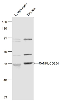
RANKL/CD254 Rabbit Polyclonal Antibody [orb11190]
FC, IF, IHC-Fr, IHC-P, WB
Canine, Rat
Human, Mouse
Rabbit
Polyclonal
Unconjugated
50 μl, 100 μl, 200 μl - Item 1 of 4


Quality Guarantee
Explore bioreagents carefree to elevate your research. All our products are rigorously tested for performance. If a product does not perform as described on its datasheet, our scientific support team will provide expert troubleshooting, a prompt replacement, or a refund. For full details, please see our Terms & Conditions and Buying Guide. Contact us at [email protected].
Protocol Information
WB
Western Blot (IB, immunoblot)
IHC
Immunohistochemistry
IF
Immunofluorescence
Available Sizes
Select a size below
Choose Conjugation or Carrier Free Version
Free Secondary Antibody (20 ul)0/0
Please add an antibody product to your cart first.