You have no items in your shopping cart.
Featured
Description
Research Area
Epigenetics
Images & Validation
−Item 1 of 2
| Tested Applications | IP, WB |
|---|---|
| Dilution Range | WB: 1-500-1-1000, IP: 1-10-1-100 |
| Reactivity | Human |
Key Properties
−| Antibody Type | Primary Antibody |
|---|---|
| Host | Rabbit |
| Clonality | Polyclonal |
| Immunogen | KLH-conjugated synthetic peptide encompassing a sequence within the center region of human LRP11. The exact sequence is proprietary. |
| Target | LRP11 |
| Purification | The antibody was purified by immunogen affinity chromatography. |
| Conjugation | Unconjugated |
Storage & Handling
−| Storage | Maintain refrigerated at 2-8°C for up to 2 weeks. For long term storage store at -20°C in small aliquots to prevent freeze-thaw cycles. |
|---|---|
| Form/Appearance | Liquid |
| Buffer/Preservatives | 0.42% Potassium phosphate, 0.87% Sodium chloride, pH 7.3, 30% glycerol, and 0.01% sodium azide. |
| Expiration Date | 12 months from date of receipt. |
| Disclaimer | For research use only |
Alternative Names
−Low-density lipoprotein receptor-related protein 11; LRP-11
Similar Products
−- Item 1 of 2
- Item 1 of 1
- Item 1 of 1
LRP11 Rabbit Polyclonal Antibody [orb185007]
WB
Human
Rabbit
Polyclonal
Unconjugated
50 μl, 100 μl, 200 μl - Item 1 of 1
LRP11 Rabbit Polyclonal Antibody [orb580987]
WB
Bovine, Canine, Equine, Guinea pig, Mouse, Porcine, Rabbit, Rat
Human
Rabbit
Polyclonal
Unconjugated
100 μl 


Quality Guarantee
Explore bioreagents carefree to elevate your research. All our products are rigorously tested for performance. If a product does not perform as described on its datasheet, our scientific support team will provide expert troubleshooting, a prompt replacement, or a refund. For full details, please see our Terms & Conditions and Buying Guide. Contact us at [email protected].

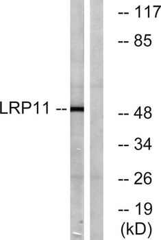
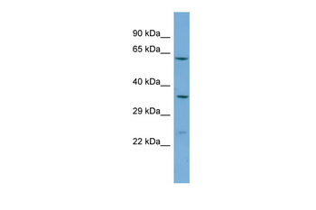
Western blot analysis of LRP11 expression in A431 (A), HepG2 (B), Jurkat (C) whole cell lysates. (Predicted band size: 53 kD; Observed band size: 49 kD)
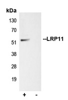
Immunoprecipitation of LRP11 from 0.5 mg Jurkat whole cell extract lysate, using 5 ug of Anti-LRP11 Antibody and 50 ul of protein G magnetic beads (+). No antibody was added to the control (-). The antibody was incubated under agitation with Protein G beads for 10 min, Jurkat whole cell extract lysate diluted in RIPA buffer was added to each sample and incubated for a further 10 min under agitation. Proteins were eluted by addition of 40 ul SDS loading buffer and incubated for 10 min at 70°C; 10 ul of each sample was separated on a SDS PAGE gel, transferred to a nitrocellulose membrane, blocked with 5% BSA and probed with Anti-LRP11 Antibody.
Documents Download
Datasheet
Product Information
Request a Document
Protocol Information
WB
Western Blot (IB, immunoblot)
IP
Immunoprecipitation
LRP11 Rabbit Polyclonal Antibody (orb215330)

- 0.0
Based on 0 reviews
Participating in our Biorbyt product reviews program enables you to support fellow scientists by sharing your firsthand experience with our products.
Login to Submit a ReviewAvailable Sizes
Select a size below
Choose Conjugation or Carrier Free Version
Free Secondary Antibody (20 ul)0/0
Please add an antibody product to your cart first.