You have no items in your shopping cart.
Description
Research Area
Disease Biomarkers, Immunology & Inflammation
Images & Validation
−Item 1 of 3
| Tested Applications | FC, IHC, WB |
|---|---|
| Dilution Range | Western blot, 0.1-0.5μg/ml Immunohistochemistry (Paraffin-embedded Section), 0.5-1μg/ml Flow Cytometry, 1-3μg/1x10^6cells |
| Reactivity | Human |
Related Conjugates & Formulations
−Key Properties
−| Antibody Type | Primary Antibody |
|---|---|
| Host | Rabbit |
| Clonality | Polyclonal |
| Isotype | Rabbit IgG |
| Immunogen | E. coli-derived human LFA3 recombinant protein (Position: F29-R215). |
| Target | Lymphocyte function-associated antigen 3 |
| Molecular Weight | 26 kDa |
| Purification | Immunogen affinity purified. |
| Conjugation | Unconjugated |
Storage & Handling
−| Storage | Maintain refrigerated at 2-8°C for up to 2 weeks. For long term storage store at -20°C in small aliquots to prevent freeze-thaw cycles. |
|---|---|
| Form/Appearance | Lyophilized |
| Buffer/Preservatives | Each vial contains antibody formulated with stabilizing components, 0.9 mg NaCl, 0.2 mg Na2HPO4, and 0.05 mg NaN3. *This antibody is supplied in a stabilized formulation. Compatibility with conjugation reactions depends on the chemistry of the conjugation method used. For conjugation methods that are not compatible with the stabilizing components present in this formulation, a carrier-free antibody format is required. |
| Concentration | Adding 0.2 ml of distilled water will yield a concentration of 500 μg/ml. |
| Expiration Date | 12 months from date of receipt. |
| Disclaimer | For research use only |
Alternative Names
−Lymphocyte function-associated antigen 3; Ag3; Surface glycoprotein LFA-3; CD58; CD58; LFA3
Similar Products
−Quality Guarantee
Explore bioreagents carefree to elevate your research. All our products are rigorously tested for performance. If a product does not perform as described on its datasheet, our scientific support team will provide expert troubleshooting, a prompt replacement, or a refund. For full details, please see our Terms & Conditions and Buying Guide. Contact us at [email protected].
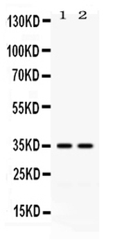
Western blot analysis of LFA3 expression in HELA whole cell lysates (lane 1) and K562 whole cell lysates (lane 2). LFA3 at 35 KD was detected using rabbit anti-LFA3 Antigen Affinity purified polyclonal antibody at 0.5 µg/mL. The blot was developed using chemiluminescence (ECL) method.
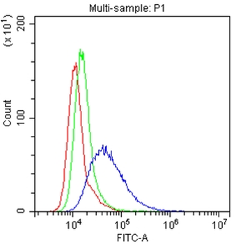
Flow Cytometry analysis of Hela cells using anti-LFA3 antibody. Overlay histogram showing Hela cells (Blue line). The cells were blocked with 10% normal goat serum. And then incubated with rabbit anti-LFA3 Antibody (1 µg/1x106 cells) for 30 min at 20°C. DyLight488 conjugated goat anti-rabbit IgG (5-10 µg/1x106 cells) was used as secondary antibody for 30 minutes at 20°C. Isotype control antibody (Green line) was rabbit IgG (1 µg/1x106) used under the same conditions. Unlabelled sample (Red line) was also used as a control.
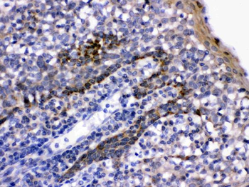
LFA3 was detected in paraffin-embedded sections of human tonsil tissues using rabbit anti-LFA3 Antigen Affinity purified polyclonal antibody at 1 µg/mL. The immunohistochemical section was developed using SABC method.
Quick Database Links
Gene Symbol
Lymphocyte function-associated antigen 3
UniProt
UniProt Details
− No UniProt data available
Documents Download
Datasheet
Product Information
Request a Document
Protocol Information
WB
Western Blot (IB, immunoblot)
IHC
Immunohistochemistry
FC
Flow Cytometry
LFA3/CD58 Rabbit Polyclonal Antibody (orb371642)

- 0.0
Based on 0 reviews
Participating in our Biorbyt product reviews program enables you to support fellow scientists by sharing your firsthand experience with our products.
Login to Submit a ReviewAvailable Sizes
Select a size below
Free Secondary Antibody (20 ul)0/0
Please add an antibody product to your cart first.