You have no items in your shopping cart.
Featured
Description
Research Area
Epigenetics
Images & Validation
−Item 1 of 2
| Tested Applications | IF, WB |
|---|---|
| Dilution Range | WB: 1:500-1:2000 |
| Reactivity | Human, Mouse, Rat |
Key Properties
−| Antibody Type | Primary Antibody |
|---|---|
| Host | Rabbit |
| Clonality | Polyclonal |
| Immunogen | Recombinant fusion protein of human HPRT |
| Target | HPRT1 |
| Purification | The antibody was purified by immunogen affinity chromatography. |
| Conjugation | Unconjugated |
Storage & Handling
−| Storage | Maintain refrigerated at 2-8°C for up to 2 weeks. For long term storage store at -20°C in small aliquots to prevent freeze-thaw cycles. |
|---|---|
| Form/Appearance | Liquid |
| Buffer/Preservatives | 0.42% Potassium phosphate, 0.87% Sodium chloride, pH 7.3, 30% glycerol, and 0.01% sodium azide. |
| Expiration Date | 12 months from date of receipt. |
| Disclaimer | For research use only |
Alternative Names
−HPRT; Hypoxanthine-guanine phosphoribosyltransferase; HGPRT; HGPRTase
Similar Products
−- Item 1 of 10
HPRT Rabbit Polyclonal Antibody [orb556857]
ICC, IHC-P, WB
Human, Mouse, Rat
Rabbit
Polyclonal
Unconjugated
100 μl - Item 1 of 7
HPRT1 Rabbit Polyclonal Antibody [orb580413]
WB
Bovine, Canine, Equine, Guinea pig, Rabbit, Rat, Zebrafish
Human, Mouse
Rabbit
Polyclonal
Unconjugated
100 μl - Item 1 of 4
- Item 1 of 4
HPRT1 Rabbit Polyclonal Antibody [orb627801]
ELISA, FC, IF, IHC, IP, WB
Human, Mouse, Rat
Rabbit
Polyclonal
Unconjugated
50 μg, 100 μg - Item 1 of 5
HPRT Rabbit Polyclonal Antibody [orb555940]
ICC, IHC-P, WB
Human, Rat, Zebrafish
Rabbit
Polyclonal
Unconjugated
100 μl


Quality Guarantee
Explore bioreagents carefree to elevate your research. All our products are rigorously tested for performance. If a product does not perform as described on its datasheet, our scientific support team will provide expert troubleshooting, a prompt replacement, or a refund. For full details, please see our Terms & Conditions and Buying Guide. Contact us at [email protected].

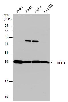
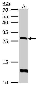
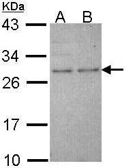
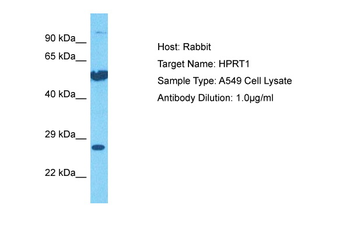
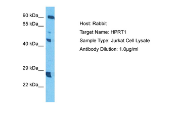
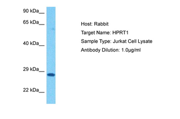
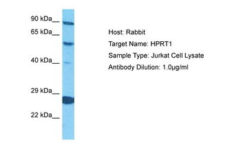
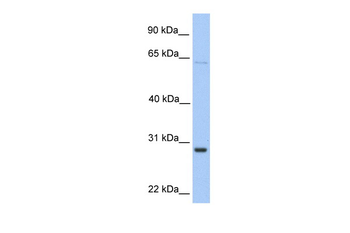
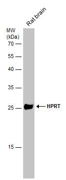
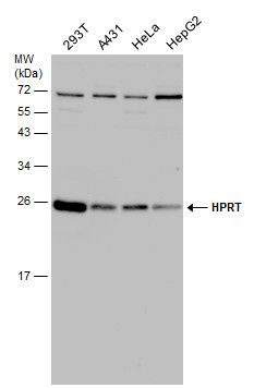
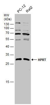
Western blot analysis of HPRT expression in SW480 (A), HT29 (B), Hela (C), mouse brain (D), mouse liver (E) whole cell lysates. (Predicted band size: 24 kD; Observed band size: 27 kD)
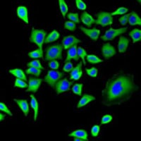
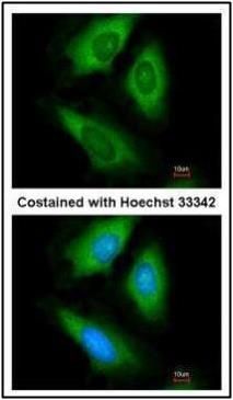
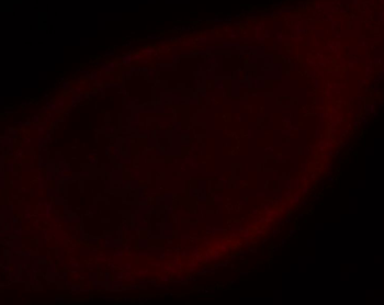
Immunofluorescent analysis of HPRT staining in U2OS cells. Formalin-fixed cells were permeabilized with 0.1% Triton X-100 in TBS for 5-10 minutes and blocked with 3% BSA-PBS for 30 minutes at room temperature. Cells were probed with the primary antibody in 3% BSA-PBS and incubated overnight at 4 °C in a humidified chamber. Cells were washed with PBST and incubated with a AF488-conjugated secondary antibody (green) in PBS at room temperature in the dark. DAPI was used to stain the cell nuclei (blue).
Documents Download
Datasheet
Product Information
Request a Document
Protocol Information
WB
Western Blot (IB, immunoblot)
IF
Immunofluorescence
HPRT Rabbit Polyclonal Antibody (orb340953)

- 0.0
Based on 0 reviews
Participating in our Biorbyt product reviews program enables you to support fellow scientists by sharing your firsthand experience with our products.
Login to Submit a ReviewAvailable Sizes
Select a size below
Choose Conjugation or Carrier Free Version
Free Secondary Antibody (20 ul)0/0
Please add an antibody product to your cart first.