You have no items in your shopping cart.
Featured
Description
Research Area
Epigenetics
Images & Validation
−Item 1 of 2
| Tested Applications | IF, WB |
|---|---|
| Dilution Range | WB: 1:500-1000 |
| Reactivity | Bovine, Gallus, Human, Monkey, Mouse, Rat |
Key Properties
−| Antibody Type | Primary Antibody |
|---|---|
| Host | Rabbit |
| Clonality | Polyclonal |
| Immunogen | KLH-conjugated synthetic peptide encompassing a sequence within the center region of human EB2. The exact sequence is proprietary. |
| Target | MAPRE2 |
| Purification | The antibody was purified by immunogen affinity chromatography. |
| Conjugation | Unconjugated |
Storage & Handling
−| Storage | Maintain refrigerated at 2-8°C for up to 2 weeks. For long term storage store at -20°C in small aliquots to prevent freeze-thaw cycles. |
|---|---|
| Form/Appearance | Liquid |
| Buffer/Preservatives | 0.42% Potassium phosphate, 0.87% Sodium chloride, pH 7.3, 30% glycerol, and 0.01% sodium azide. |
| Expiration Date | 12 months from date of receipt. |
| Disclaimer | For research use only |
Alternative Names
−RP1; Microtubule-associated protein RP/EB family member 2; APC-binding protein EB2; End-binding protein 2; EB2
Similar Products
−- Item 1 of 2
EB2 Rabbit Polyclonal Antibody [orb101410]
IF, IHC-Fr, IHC-P, WB
Bovine, Canine, Equine, Rat
Human, Mouse
Rabbit
Polyclonal
Unconjugated
50 μl, 200 μl, 100 μl - Item 1 of 1
MAPRE2 Antibody (N-term) [orb1935505]
WB
Mouse, Other, Rat
Human
Rabbit
Polyclonal
Unconjugated
50 μl, 100 μl - Item 1 of 1
MAPRE2 Rabbit Polyclonal Antibody [orb586626]
WB
Bovine, Canine, Equine, Guinea pig, Mouse, Rabbit, Rat, Yeast
Human
Rabbit
Polyclonal
Unconjugated
100 μl - Item 1 of 1
MAPRE2 Rabbit Polyclonal Antibody [orb586627]
WB
Bovine, Canine, Equine, Guinea pig, Mouse, Rabbit, Rat
Human
Rabbit
Polyclonal
Unconjugated
100 μl 
MAPRE2 Polyclonal Antibody [orb3150545]
ELISA, IHC, WB
Bovine, Frog, Gallus, Human, Mouse, Rat
Rabbit
Polyclonal
Unconjugated
50 μg, 100 μg
Quality Guarantee
Explore bioreagents carefree to elevate your research. All our products are rigorously tested for performance. If a product does not perform as described on its datasheet, our scientific support team will provide expert troubleshooting, a prompt replacement, or a refund. For full details, please see our Terms & Conditions and Buying Guide. Contact us at [email protected].
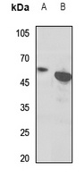
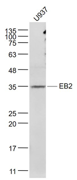
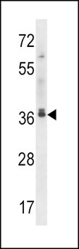
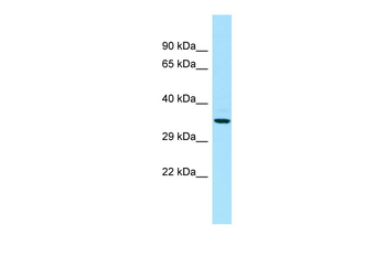
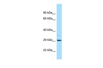
Western blot analysis of EB2 expression in mouse heart (A), rat heart (B) whole cell lysates. (Predicted band size: 37 kD; Observed band size: 50 kD)
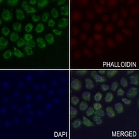
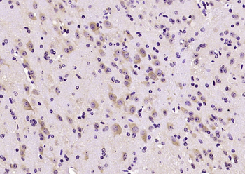
Immunofluorescent analysis of EB2 staining in SGC7901 cells. Formalin-fixed cells were permeabilized with 0.1% Triton X-100 in TBS for 5-10 minutes and blocked with 3% BSA-PBS for 30 minutes at room temperature. Cells were probed with the primary antibody in 3% BSA-PBS and incubated overnight at 4 °C in a hidified chamber. Cells were washed with PBST and incubated with a AF488-conjugated secondary antibody (green) in PBS at room temperature in the dark. Phalloidin - AF594 was used to stain Actin filaments (red). DAPI was used to stain the cell nuclei (blue).
Documents Download
Datasheet
Product Information
Request a Document
Protocol Information
WB
Western Blot (IB, immunoblot)
IF
Immunofluorescence
Filter by Applications
Filter by Species
Stefano Pezzini 1, Aurora Mustaccia 2, Pierre Aboa 1, Giorgia Faustini 1, Alessio Branchini 3, Mirko Pinotti 3, Angelisa Frasca 1, Joseph J Porter 4, John D Lueck 4, Nicoletta Landsberger Engineered tRNAs efficiently suppress CDKL5 premature termination codons Engineered tRNAs efficiently suppress CDKL5 premature termination codons, Sci Rep, (2024)
Applications
WB
Reactivity
Human
EB2 Rabbit Polyclonal Antibody (orb234827)

- 0.0
Based on 0 reviews
Participating in our Biorbyt product reviews program enables you to support fellow scientists by sharing your firsthand experience with our products.
Login to Submit a ReviewAvailable Sizes
Select a size below
Choose Conjugation or Carrier Free Version
Free Secondary Antibody (20 ul)0/0
Please add an antibody product to your cart first.