You have no items in your shopping cart.
Description
Research Area
Cell Biology, Immunology & Inflammation, Protein Biochemistry, Signal Transduction
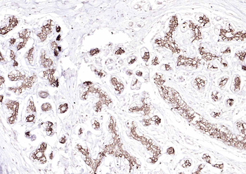
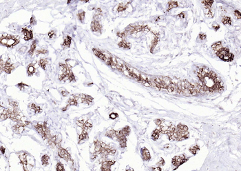
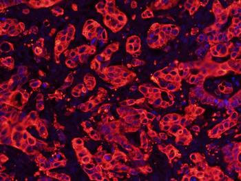
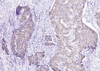
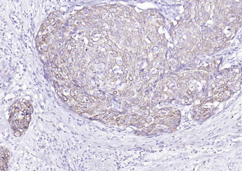
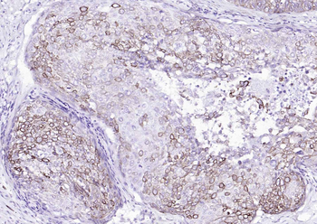
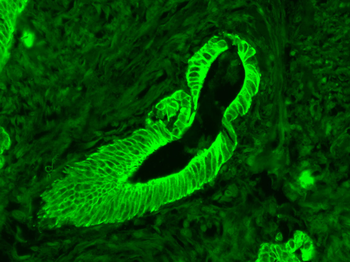
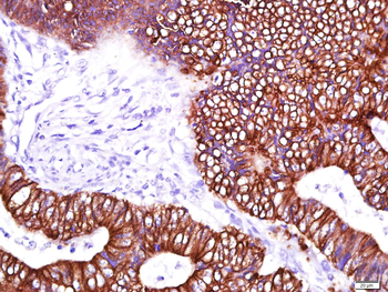
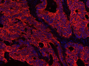
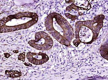
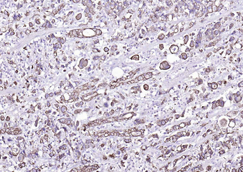
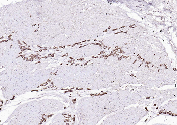
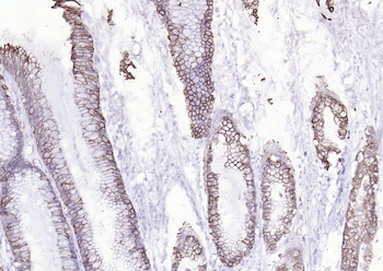
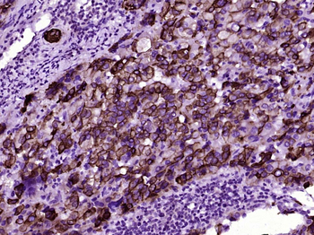
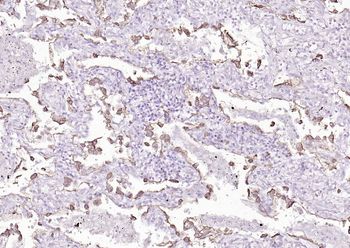
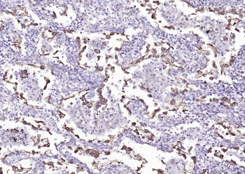
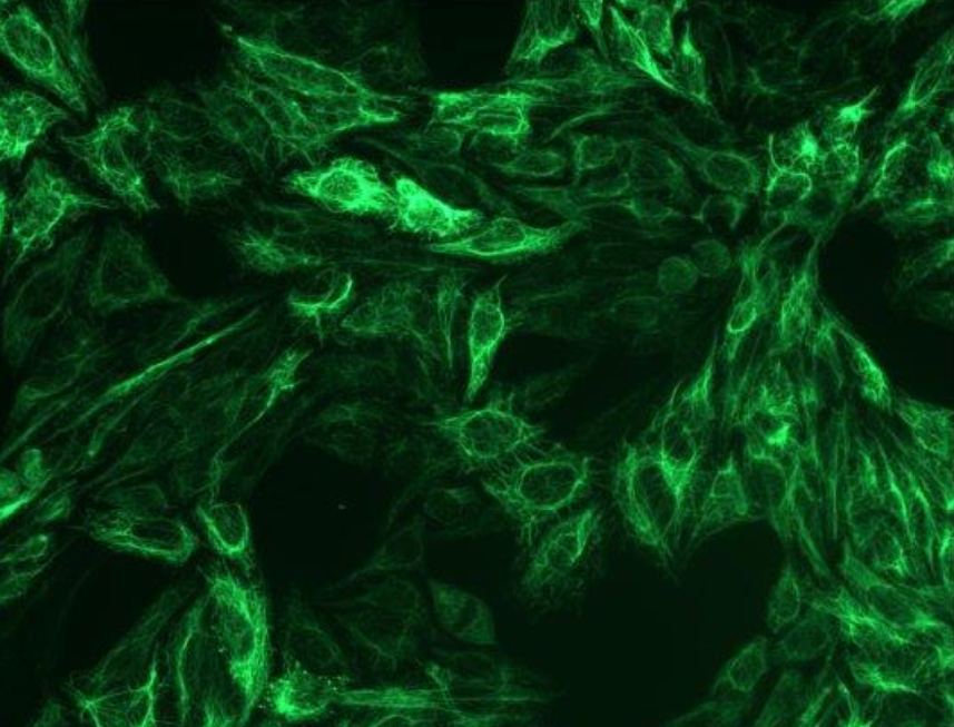
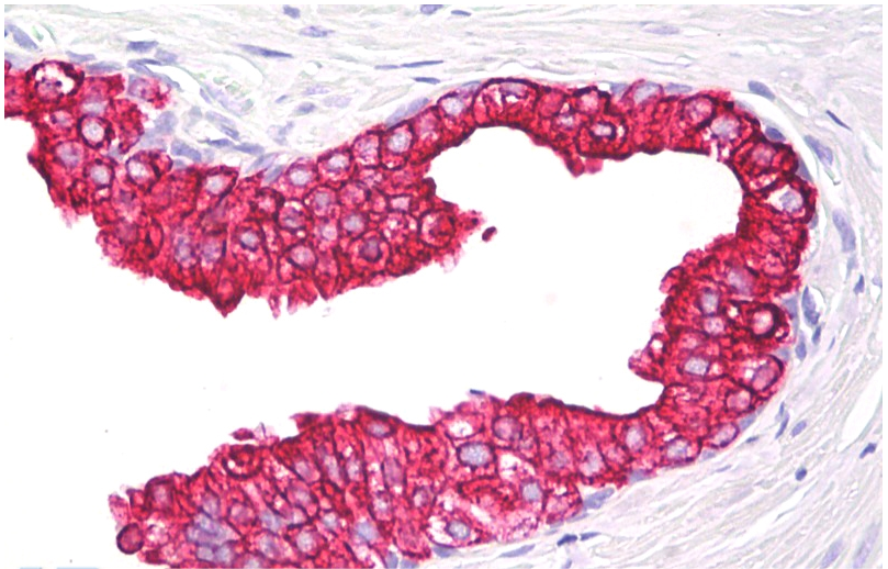
Images & Validation
−
Item 1 of 6
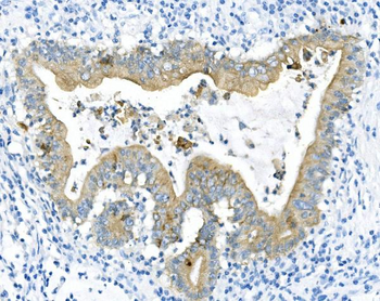
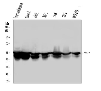
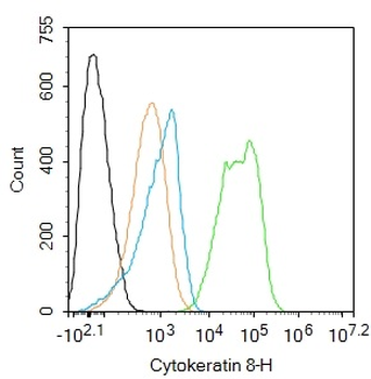
| Tested Applications | FC, ICC, IF, IHC, IHC-Fr, WB |
|---|---|
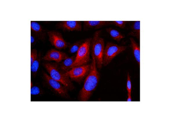
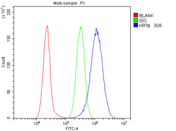
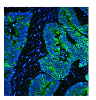
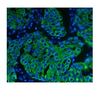
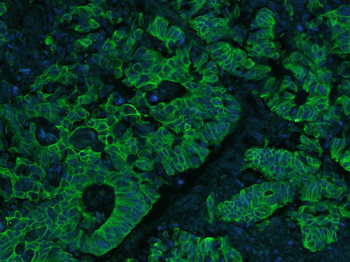
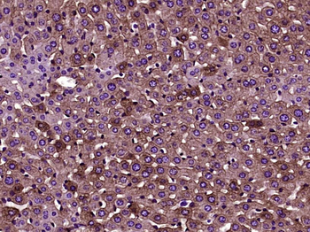
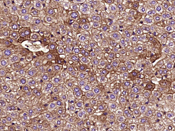
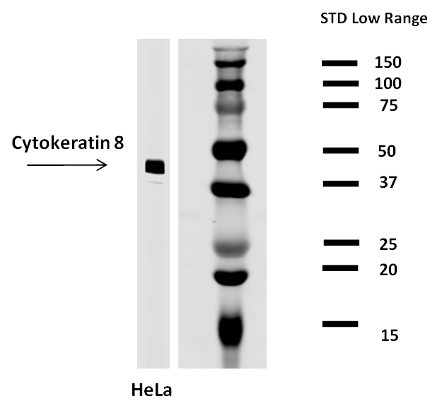
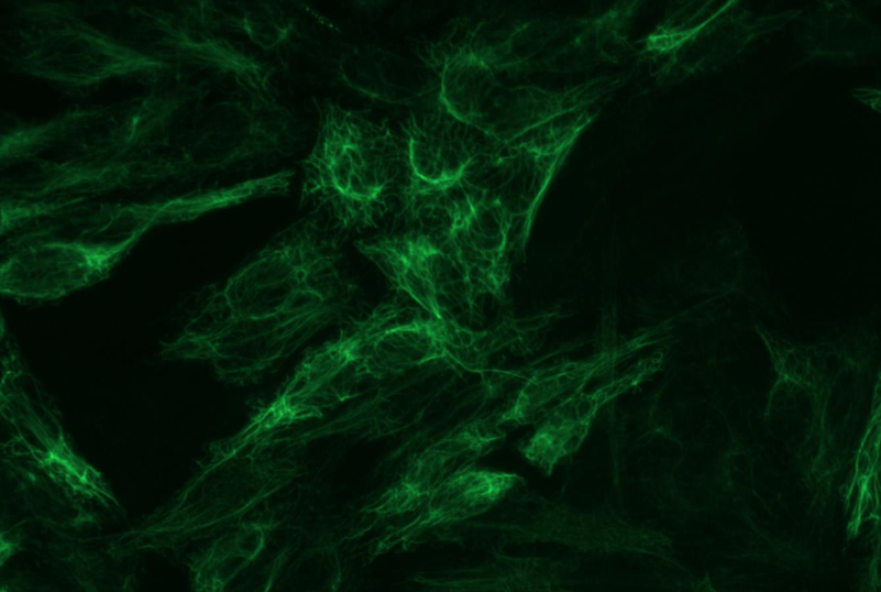
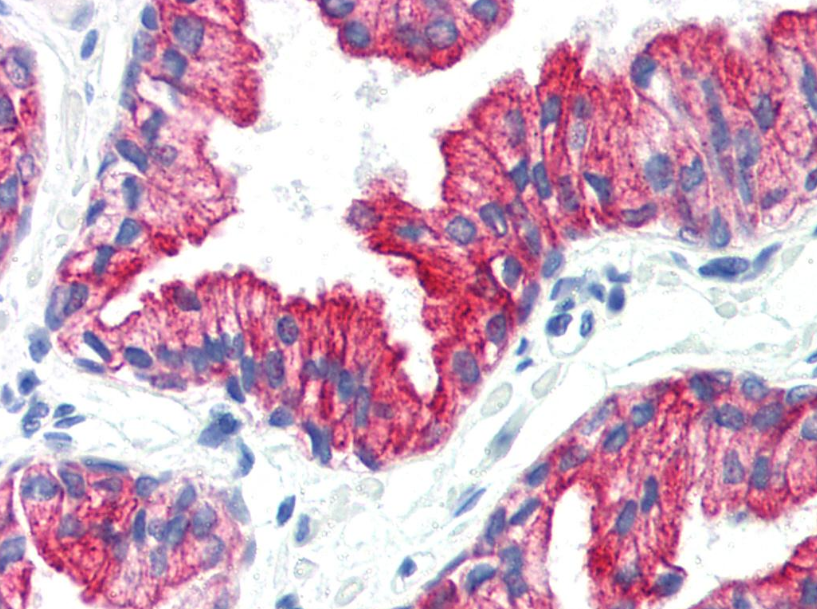
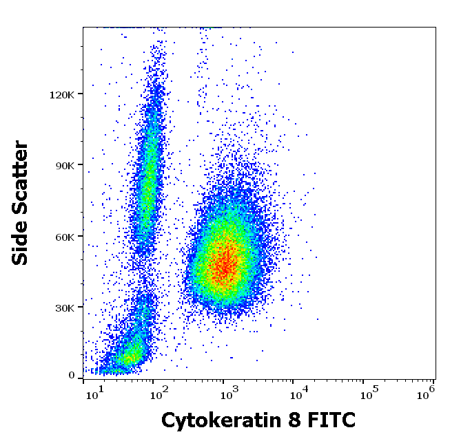
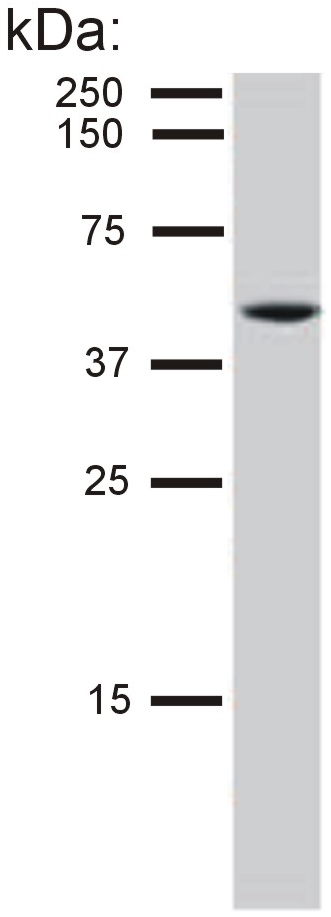
| Dilution Range | Western blot, 0.1-0.5μg/ml Immunohistochemistry (Paraffin-embedded Section), 0.5-1μg/ml Immunohistochemistry (Frozen Section), 0.5-1μg/ml Immunofluorescence, 2μg/ml Immunocytochemistry/Immunofluorescence, 2μg/ml Flow Cytometry (Fixed), 1-3μg/1x10^6 cells |
| Reactivity | Human, Mouse, Rat |
Related Conjugates & Formulations
−Key Properties
−| Antibody Type | Primary Antibody |
|---|---|
| Host | Mouse |
| Clonality | Monoclonal |
| Isotype | Mouse IgG2b |
| Clone No. | B5I1 |
| Immunogen | E.coli-derived human Cytokeratin 8 recombinant protein (Position: D107-K325). Human Cytokeratin 8 shares 95.4% and 94.5% amino acid (aa) sequence identity with mouse and rat Cytokeratin 8, respectively. |
| Target | Keratin, type II cytoskeletal 8 |
| Molecular Weight | 54 kDa |
| Purification | Immunogen affinity purified. |
| Conjugation | Unconjugated |
Storage & Handling
−| Storage | Maintain refrigerated at 2-8°C for up to 2 weeks. For long term storage store at -20°C in small aliquots to prevent freeze-thaw cycles. |
|---|---|
| Form/Appearance | Lyophilized |
| Buffer/Preservatives | Each vial contains 4mg Trehalose, 0.9mg NaCl, 0.2mg Na2HPO4, 0.05mg NaN3. |
| Concentration | Adding 0.2 ml of distilled water will yield a concentration of 500 μg/ml. |
| Expiration Date | 12 months from date of receipt. |
| Disclaimer | For research use only |
Alternative Names
−CARD2; Cell and organelle markers; CK 8; CK8; CYK8; Cytokeratin 8; Cytoskeleton Marker; K2C8; K8; keratin 8; KO; KRT8; Type II keratin Kb8
Similar Products
−- Item 1 of 22
Cytokeratin 8 Mouse Monoclonal Antibody [orb500822]
FC, ICC, IF, IHC-Fr, IHC-P
Mouse, Rat
Human, Mouse, Rat
Mouse
Monoclonal
Unconjugated
50 μl, 100 μl, 200 μl, 200 μg - Item 1 of 3
Cytokeratin 8 Antibody [orb43712]
FC, ICC, IHC-P, IP, WB
Bovine, Human, Porcine, Rabbit, Sheep
Monoclonal
Unconjugated
0.1 mg - Item 1 of 2
Cytokeratin 8 Antibody (FITC) [orb154469]
FC
Bovine, Human, Porcine, Rabbit, Sheep
Monoclonal
FITC
0.1 mg - Item 1 of 2
Cytokeratin 8 Antibody [orb43716]
ICC, IHC-P, IP, WB
Bovine, Human, Porcine, Sheep
Monoclonal
Unconjugated
0.1 mg - Item 1 of 1
Cytokeratin 5/8 Antibody [orb43715]
ICC, IHC-P, IP, WB
Bovine, Canine, Hamster, Human, Mouse, Porcine, Rat, Sheep
Monoclonal
Unconjugated
0.1 mg


Quality Guarantee
Explore bioreagents carefree to elevate your research. All our products are rigorously tested for performance. If a product does not perform as described on its datasheet, our scientific support team will provide expert troubleshooting, a prompt replacement, or a refund. For full details, please see our Terms & Conditions and Buying Guide. Contact us at [email protected].
Quick Database Links
Gene Symbol
Keratin, type II cytoskeletal 8
UniProt
UniProt Details
− No UniProt data available
Protocol Information
WB
Western Blot (IB, immunoblot)
IHC
Immunohistochemistry
IHC-Fr
Immunohistochemistry Frozen
FC
Flow Cytometry
IF
Immunofluorescence
ICC
Immunocytochemistry
Available Sizes
Select a size below
Free Secondary Antibody (20 ul)0/0
Please add an antibody product to your cart first.