You have no items in your shopping cart.
Description
Research Area
Epigenetics, Infectious Diseases
Images & Validation
−Item 1 of 4
| Tested Applications | ELISA, FC, ICC, IHC-P, IP, WB |
|---|---|
| Reactivity | Human |
| Application Notes |
Key Properties
−| Antibody Type | Primary Antibody |
|---|---|
| Clonality | Monoclonal |
| Isotype | Mouse IgG1 |
| Clone No. | DC-10 |
| Immunogen | Human breast carcinoma cell line PMC-42. |
| Target | Cytokeratin 18 |
| Purification | Purified by protein-A affinity chromatography. |
| Conjugation | Unconjugated |
Storage & Handling
−| Storage | Maintain refrigerated at 2-8°C for up to 2 weeks. For long term storage store at -20°C in small aliquots to prevent freeze-thaw cycles. |
|---|---|
| Buffer/Preservatives | Phosphate buffered saline (PBS), pH 7.4, 15 mM sodium azide |
| Concentration | 1 mg/ml |
| Expiration Date | 12 months from date of receipt. |
| Disclaimer | For research use only |
Alternative Names
−K18, CK18, CYK18, KRT18
Similar Products
−- Item 1 of 21
Cytokeratin 18/KRT18 Rabbit Polyclonal Antibody [orb389489]
FC, IF, IHC, WB
Human, Mouse, Rat
Rabbit
Polyclonal
Unconjugated
100 μg - Item 1 of 12
CK18 Mouse Monoclonal Antibody [orb499665]
FC, ICC, IF, IHC-Fr, IHC-P, WB
Mouse, Rat
Human
Mouse
Monoclonal
Unconjugated
100 μl, 200 μl, 200 μg, 50 μl - Item 1 of 10
CK18 Recombinant Rabbit Monoclonal Antibody [orb500277]
ICC, IF, IHC-Fr, IHC-P, WB
Mouse, Rat
Human, Mouse, Rat
Rabbit
Recombinant
Unconjugated
50 μl, 100 μl - Item 1 of 8
CK18 Rabbit Polyclonal Antibody [orb5871]
FC, ICC, IF, IHC-Fr, IHC-P, KO/KD Validated, WB
Canine, Gallus, Rabbit
Human, Mouse, Rat
Rabbit
Polyclonal
Unconjugated
50 μl, 100 μl, 200 μl - Item 1 of 6
Mouse Cytokeratin 18 / Keratin K18 Antibody [orb98259]
FC, ICC, IHC-Fr, WB
Canine, Gallus, Hamster, Human, Mouse, Porcine, Rabbit, Rat, Zebrafish
Mouse
Monoclonal
Unconjugated
0.1 mg


Quality Guarantee
Explore bioreagents carefree to elevate your research. All our products are rigorously tested for performance. If a product does not perform as described on its datasheet, our scientific support team will provide expert troubleshooting, a prompt replacement, or a refund. For full details, please see our Terms & Conditions and Buying Guide. Contact us at [email protected].
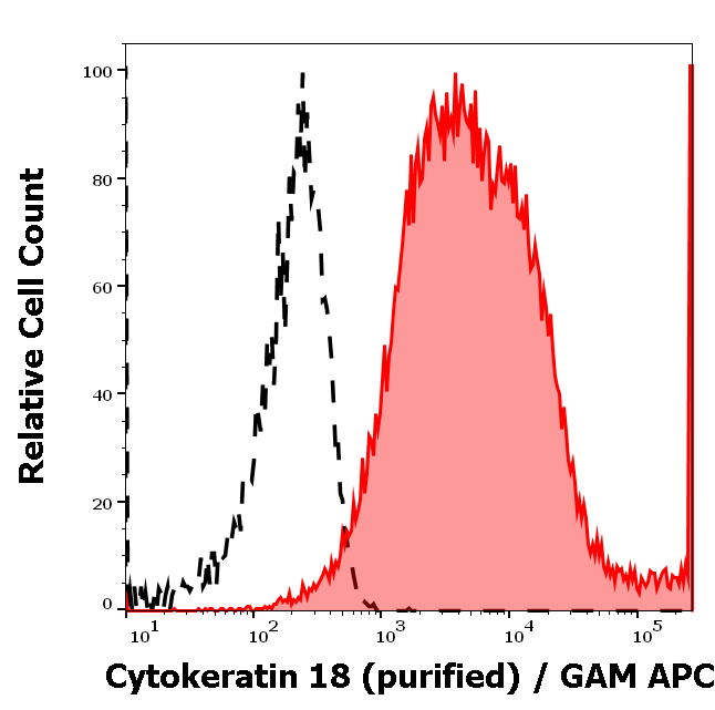
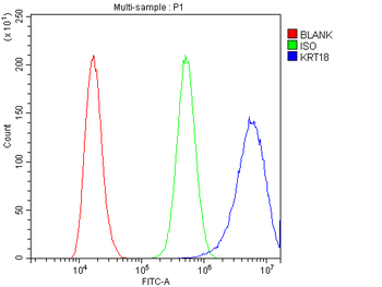
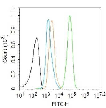
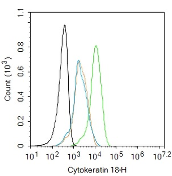
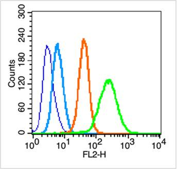
Separation of MCF-7 cells (red-filled) from Caco-2 cells (black-dashed) in flow cytometry analysis (intracellular staining) of cells stained using anti-cytokeratin 18 (DC-10) purified antibody (concentration in sample 0.6 μg/ml, GAM APC).
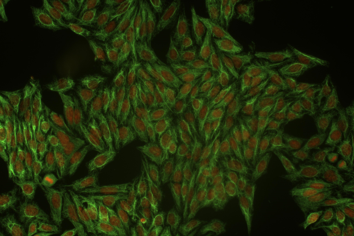
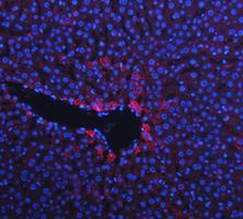
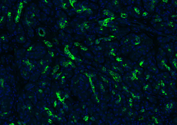
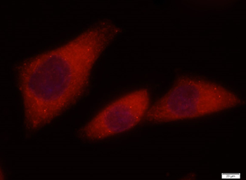
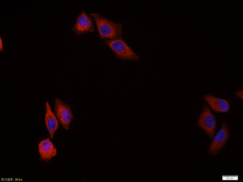
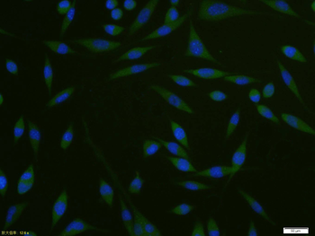
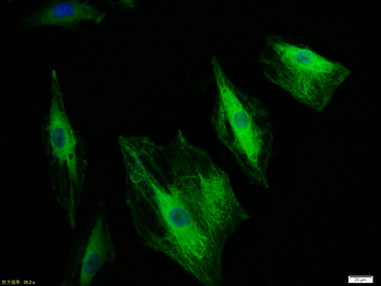
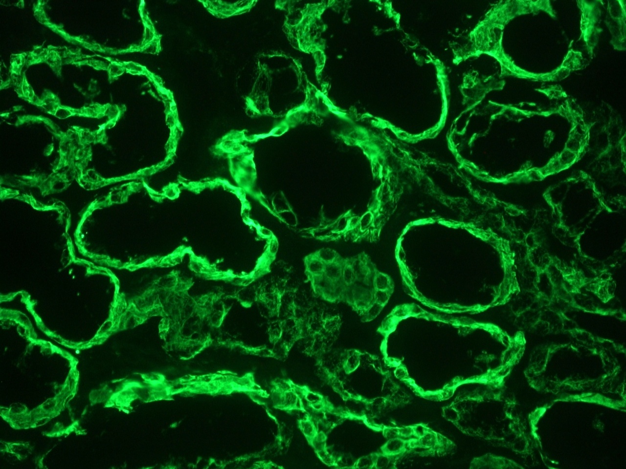
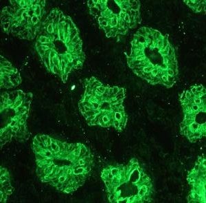
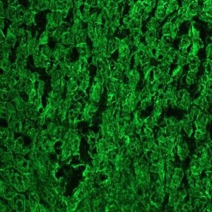
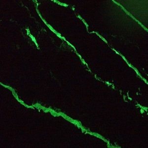
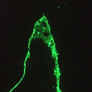
Immunocytochemistry staining of cytokeratin 18 in Hep-2 cells using mouse monoclonal antibody DC-10 (orb43750, diluted 1:400), detected with GAM IgG-Alexa Fluor®488 (diluted 1:200; green), cell nuclei stained with PI (1 µg/ml; orange).
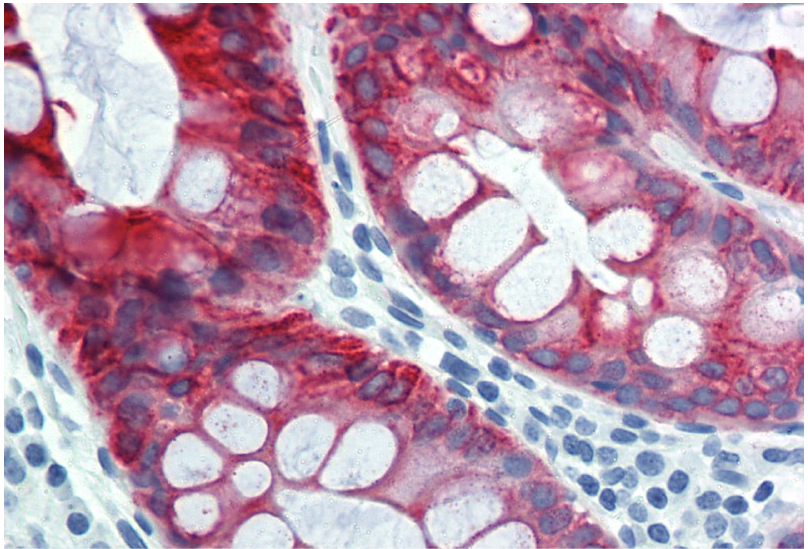
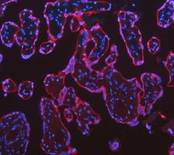
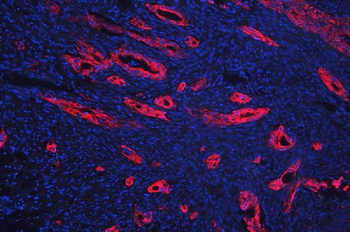
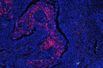
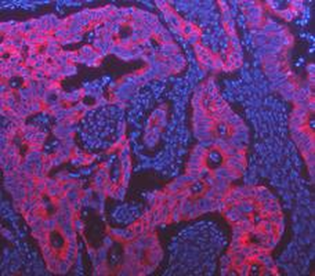
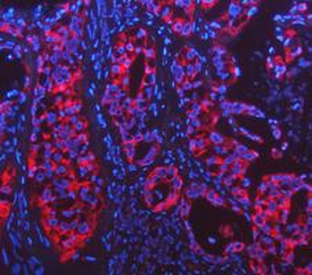
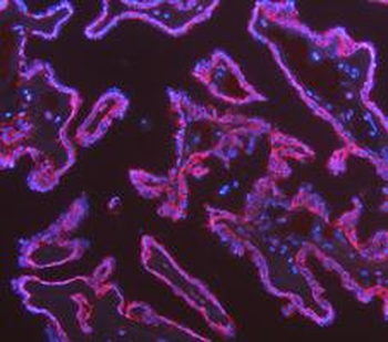
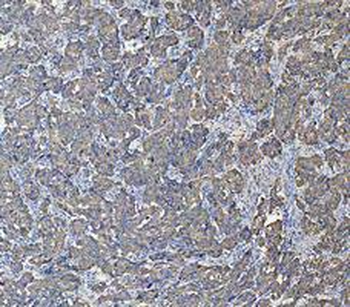
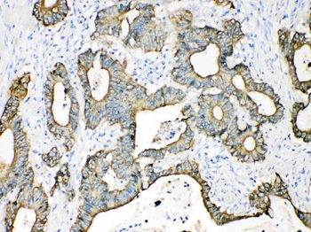
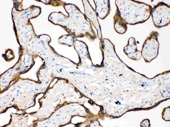
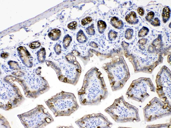
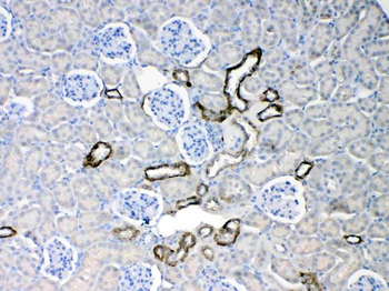
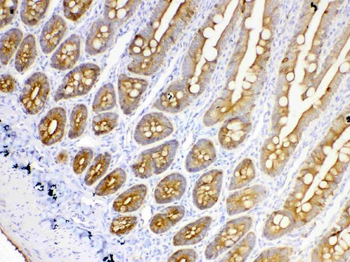
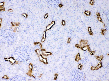
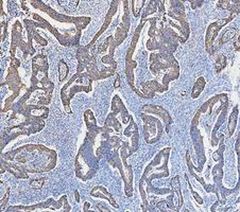
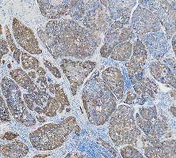
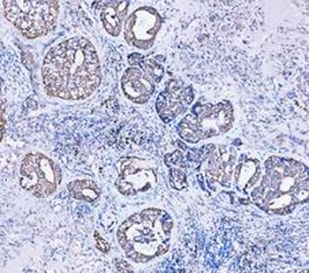
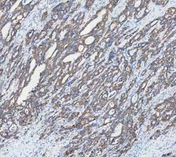
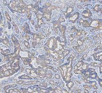
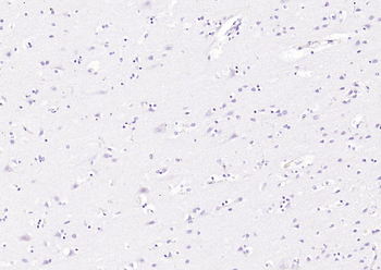
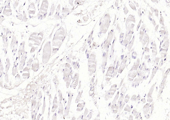
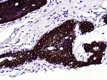
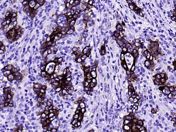
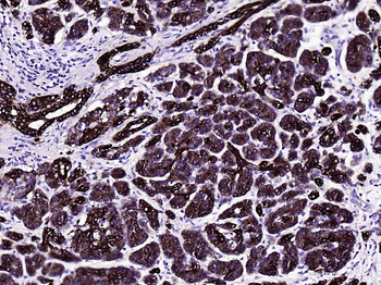
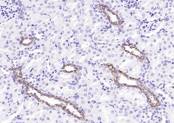
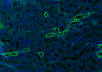
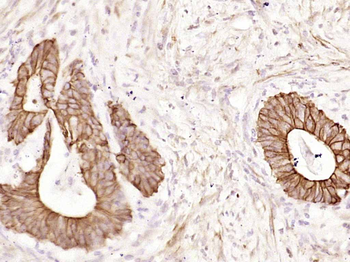
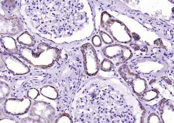
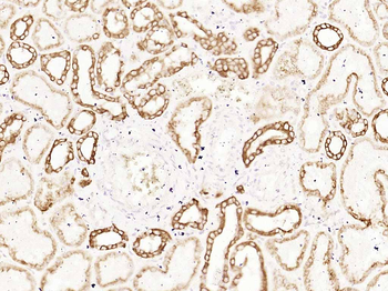
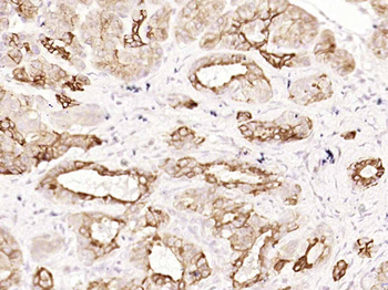
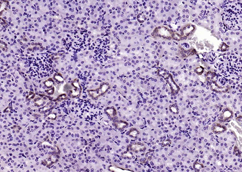
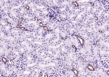
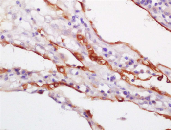
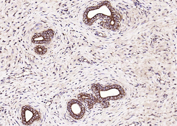
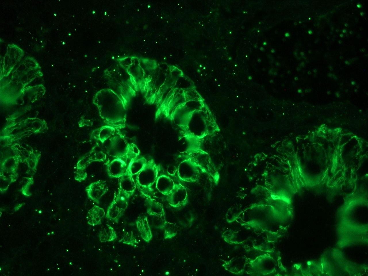
Immunohistochemistry staining of human colon (paraffin sections) using anti-cytokeratin 18 (clone DC-10).
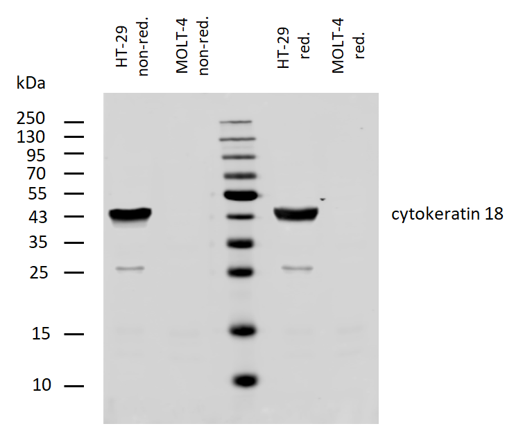
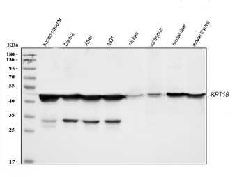
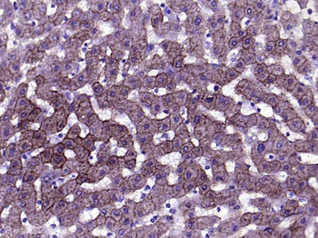
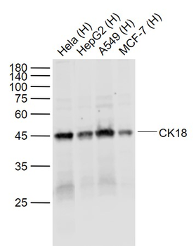
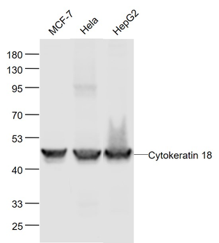
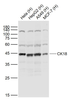
Western blotting analysis of human cytokeratin 18 using mouse monoclonal antibody DC-10 on lysates of HT-29 cell line and MOLT-4 cell line (cytokeratin non-expressing cell line; negative control) under non-reducing and reducing conditions. Nitrocellulose membrane was probed with 2 µg/ml of mouse anti-cytokeratin 18 monoclonal antibody followed by IRDye800-conjugated anti-mouse secondary antibody. A specific band was detected for cytokeratin 18 at approximately 46 kDa, and its proteolytic fragment at approximately 25 kDa.
Documents Download
Datasheet
Product Information
Request a Document
Protocol Information
WB
Western Blot (IB, immunoblot)
IHC-P
Immunohistochemistry Paraffin
FC
Flow Cytometry
ICC
Immunocytochemistry
ELISA
Enzyme-linked Immunosorbent Assay (EIA)
IP
Immunoprecipitation
Cytokeratin 18 Antibody (orb43750)

- 0.0
Based on 0 reviews
Participating in our Biorbyt product reviews program enables you to support fellow scientists by sharing your firsthand experience with our products.
Login to Submit a ReviewAvailable Sizes
Select a size below
Free Secondary Antibody (20 ul)0/0
Please add an antibody product to your cart first.