You have no items in your shopping cart.
Featured
Description
Images & Validation
−Item 1 of 5
| Tested Applications | IF, IHC, WB |
|---|---|
| Dilution Range | WB: 1-500-1-1000, IHC-P: 1-100-1-200 |
| Reactivity | Bovine, Human, Mouse, Porcine, Rat |
Key Properties
−| Antibody Type | Primary Antibody |
|---|---|
| Host | Rabbit |
| Clonality | Polyclonal |
| Immunogen | KLH-conjugated synthetic peptide encompassing a sequence within the center region of human Cyclin G1. The exact sequence is proprietary. |
| Target | CCNG1 |
| Molecular Weight | 34 kDa |
| Purification | The antibody was purified by immunogen affinity chromatography. |
| Conjugation | Unconjugated |
Storage & Handling
−| Storage | Maintain refrigerated at 2-8°C for up to 2 weeks. For long term storage store at -20°C in small aliquots to prevent freeze-thaw cycles. |
|---|---|
| Form/Appearance | Liquid |
| Buffer/Preservatives | 0.42% Potassium phosphate, 0.87% Sodium chloride, pH 7.3, 30% glycerol, and 0.01% sodium azide. |
| Expiration Date | 12 months from date of receipt. |
| Disclaimer | For research use only |
Alternative Names
−CCNG; CYCG1; Cyclin-G1; Cyclin-G
Similar Products
−- Item 1 of 9
Cyclin D1 Rabbit Polyclonal Antibody [orb10496]
FC, ICC, IF, IHC-Fr, IHC-P, WB
Canine
Human, Mouse, Rat
Rabbit
Polyclonal
Unconjugated
50 μl, 100 μl, 200 μl - Item 1 of 10
Cyclin D1 Rabbit Polyclonal Antibody [orb500813]
FC, ICC, IF, IHC-Fr, IHC-P
Mouse, Rat
Human, Mouse, Rat
Rabbit
Polyclonal
Unconjugated
50 μl, 100 μl, 200 μl - Item 1 of 7
CDK1 Rabbit Polyclonal Antibody [orb10355]
ICC, IF, IHC-Fr, IHC-P, WB
Canine, Equine, Rat
Human, Mouse
Rabbit
Polyclonal
Unconjugated
50 μl, 100 μl, 200 μl - Item 1 of 7
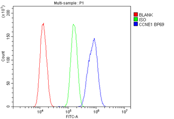
Cyclin E1/CCNE1 Rabbit Polyclonal Antibody [orb654316]
ELISA, FC, IHC, WB
Human, Mouse, Rat
Rabbit
Polyclonal
Unconjugated
100 μg - Item 1 of 6
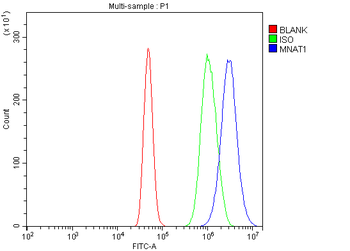
MNAT1 Rabbit Polyclonal Antibody [orb312140]
FC, ICC, IF, IHC, WB
Human, Mouse, Rat
Rabbit
Polyclonal
Unconjugated
100 μg
Quality Guarantee
Explore bioreagents carefree to elevate your research. All our products are rigorously tested for performance. If a product does not perform as described on its datasheet, our scientific support team will provide expert troubleshooting, a prompt replacement, or a refund. For full details, please see our Terms & Conditions and Buying Guide. Contact us at [email protected].
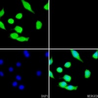
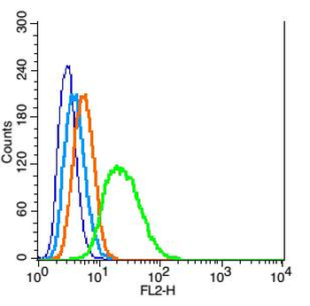
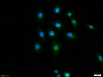
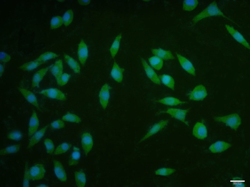
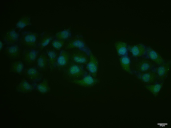
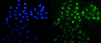
Immunofluorescent analysis of Cyclin G1 staining in B12 cells. Formalin-fixed cells were permeabilized with 0.1% Triton X-100 in TBS for 5-10 minutes and blocked with 3% BSA-PBS for 30 minutes at room temperature. Cells were probed with the primary antibody in 3% BSA-PBS and incubated overnight at 4 °C in a hidified chamber. Cells were washed with PBST and incubated with a AF488-conjugated secondary antibody (green) in PBS at room temperature in the dark. Phalloidin - AF594 was used to stain Actin filaments (red). DAPI was used to stain the cell nuclei (blue).
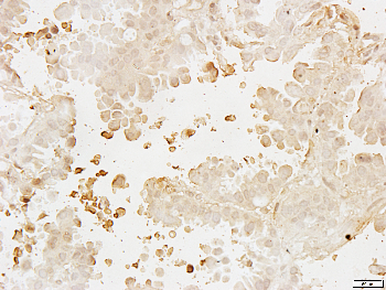
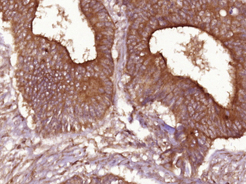
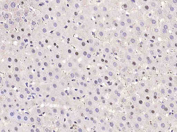
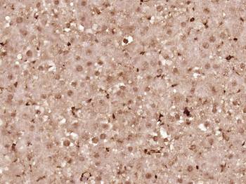
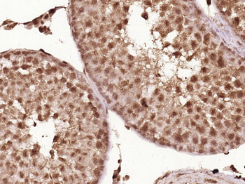
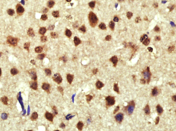
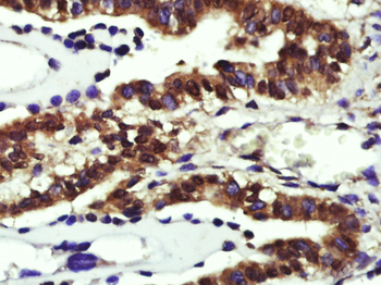
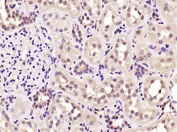
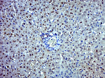
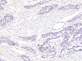
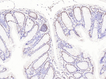
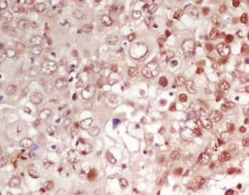
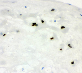
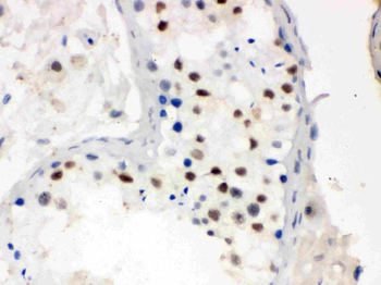
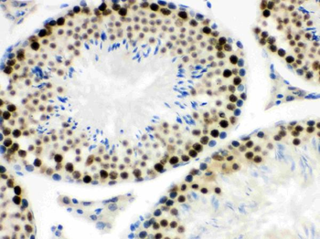
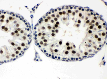
Immunohistochemical analysis of paraffin-embedded human lung cancer using Cyclin G1 Rabbit Polyclonal Antibody orb213680 with dilution at 1:100.
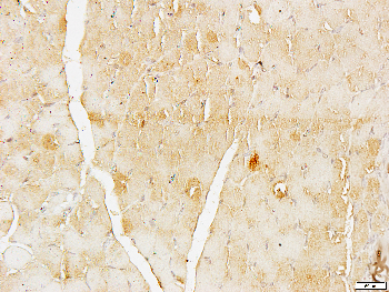
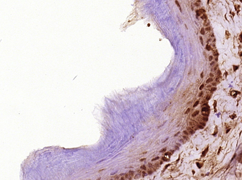
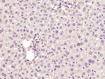
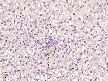
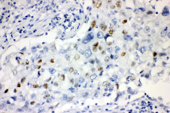
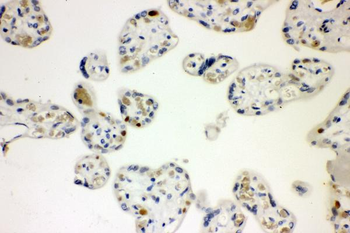
Immunohistochemical analysis of paraffin-embedded mouse muscle using Cyclin G1 Rabbit Polyclonal Antibody orb213680 with dilution at 1:100.
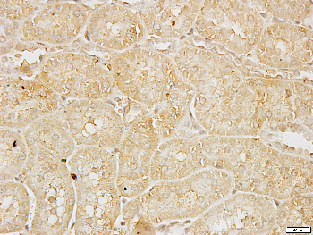
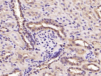
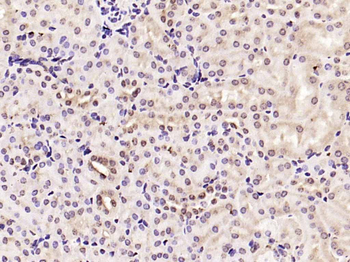
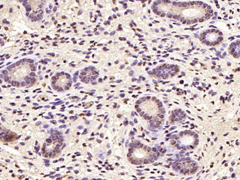
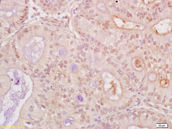
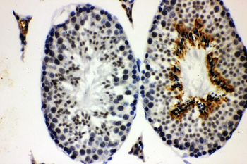
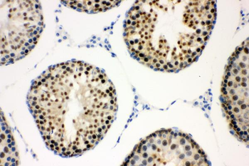
Immunohistochemical analysis of paraffin-embedded rat stomach using Cyclin G1 Rabbit Polyclonal Antibody orb213680 with dilution at 1:100.
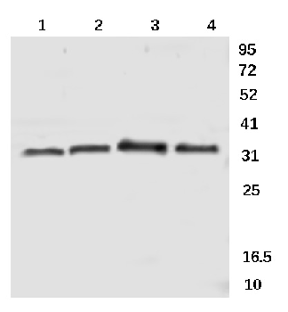
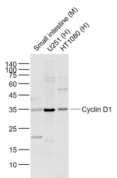
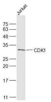
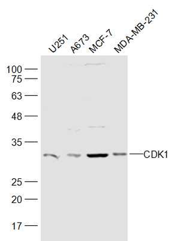
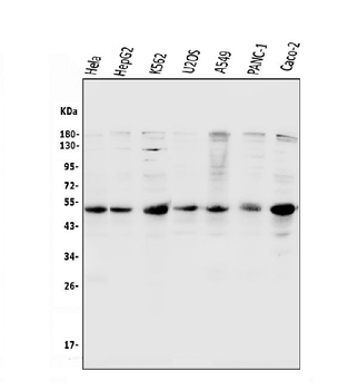
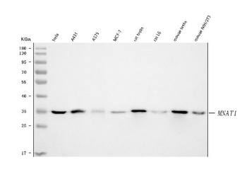
Western blot analysis of rat kidney (1), mouse kidney (2), rat heart (3), mouse heart (4) using Cyclin G1 Rabbit Polyclonal Antibody orb213680 with dilution at 1:1000.
Documents Download
Datasheet
Product Information
Request a Document
Protocol Information
WB
Western Blot (IB, immunoblot)
IHC
Immunohistochemistry
IF
Immunofluorescence
Aguilera-Rojas, Matias et al. Deregulation of miR-27a may contribute to canine fibroblast activation after coculture with a mast cell tumour cell line FEBS Open Bio, 10, 802-816 (2020)
Cyclin G1 Rabbit Polyclonal Antibody (orb213680)

- 0.0
Based on 0 reviews
Participating in our Biorbyt product reviews program enables you to support fellow scientists by sharing your firsthand experience with our products.
Login to Submit a ReviewAvailable Sizes
Select a size below
Choose Conjugation or Carrier Free Version
Free Secondary Antibody (20 ul)0/0
Please add an antibody product to your cart first.