You have no items in your shopping cart.
Featured
Description
Research Area
Apoptotic, Cancer, Epigenetics, Immunology, Neuroscience, Signaling Pathways
Images & Validation
−Item 1 of 3
| Tested Applications | IF, IHC, WB |
|---|---|
| Dilution Range | WB: 1:500-1000, IHC-P: 1:100-200, IF/ICC: 1:100-500 |
| Reactivity | Human, Mouse, Rat |
Key Properties
−| Antibody Type | Primary Antibody |
|---|---|
| Host | Rabbit |
| Clonality | Polyclonal |
| Immunogen | KLH-conjugated synthetic peptide encompassing a sequence within the N-term region of human ASC. The exact sequence is proprietary. |
| Target | PYCARD |
| Purification | The antibody was purified by immunogen affinity chromatography. |
| Conjugation | Unconjugated |
Storage & Handling
−| Storage | Maintain refrigerated at 2-8°C for up to 2 weeks. For long term storage store at -20°C in small aliquots to prevent freeze-thaw cycles. |
|---|---|
| Form/Appearance | Liquid |
| Buffer/Preservatives | 0.42% Potassium phosphate, 0.87% Sodium chloride, pH 7.3, 30% glycerol, and 0.01% sodium azide. |
| Expiration Date | 12 months from date of receipt. |
| Disclaimer | For research use only |
Alternative Names
−ASC; CARD5; TMS1; Apoptosis-associated speck-like protein containing a CARD; hASC; Caspase recruitment domain-containing protein 5; PYD and CARD domain-containing protein; Target of methylation-induced silencing 1
Similar Products
−- Item 1 of 12
ASC/TMS1/PYCARD Rabbit Polyclonal Antibody [orb1728091]
ELISA, FC, ICC, IF, IHC, WB
Human
Rabbit
Polyclonal
Unconjugated
100 μg - Item 1 of 7
ASC/TMS1 Rabbit Polyclonal Antibody [orb100371]
WB
Human
Rabbit
Polyclonal
Unconjugated
50 μl, 100 μl, 200 μl - Item 1 of 3
Steroid sulfatase/STS Rabbit Polyclonal Antibody [orb763085]
ELISA, FC, IHC, WB
Human
Rabbit
Polyclonal
Unconjugated
100 μg - Item 1 of 3
TMS1/ASC/Pycard Rabbit Polyclonal Antibody [orb745962]
ELISA, FC, IHC, WB
Mouse, Rat
Rabbit
Polyclonal
Unconjugated
100 μg - Item 1 of 3
SLC1A5 Rabbit Polyclonal Antibody [orb526711]
WB
Human
Human, Mouse
Rabbit
Polyclonal
Unconjugated
50 μl, 100 μl, 200 μl


Quality Guarantee
Explore bioreagents carefree to elevate your research. All our products are rigorously tested for performance. If a product does not perform as described on its datasheet, our scientific support team will provide expert troubleshooting, a prompt replacement, or a refund. For full details, please see our Terms & Conditions and Buying Guide. Contact us at [email protected].
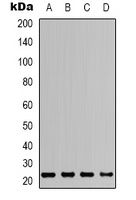
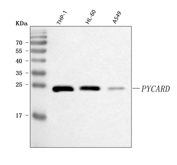
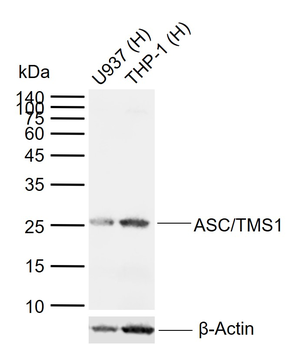
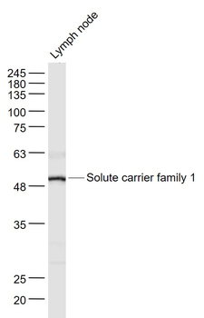
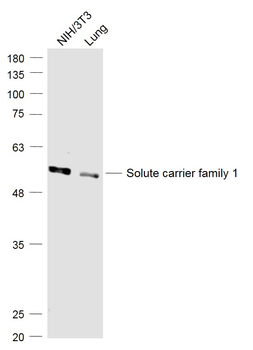
Western blot analysis of ASC expression in HEK293T (A), Raw264.7 (B), mouse brain (C), rat brain (D) whole cell lysates. (Predicted band size: 21 kD; Observed band size: 24 kD)
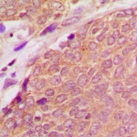
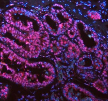
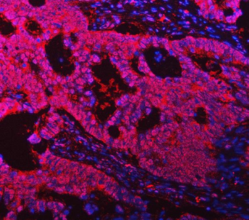
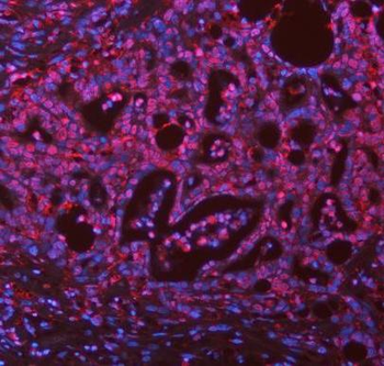
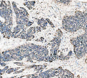
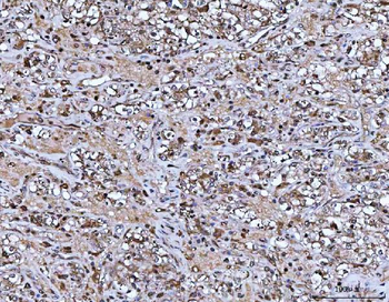
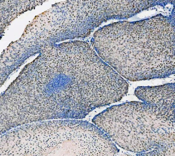
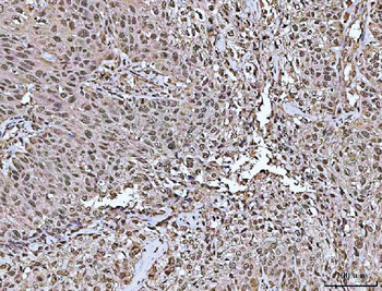
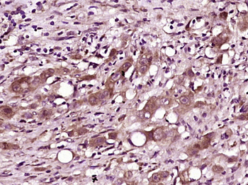
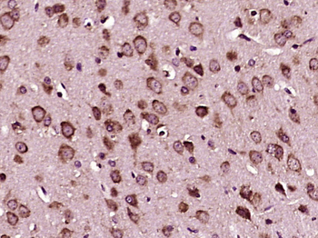
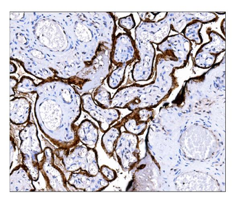
Immunohistochemical analysis of ASC staining in human breast cancer formalin fixed paraffin embedded tissue section. The section was pre-treated using heat mediated antigen retrieval with sodium citrate buffer (pH 6.0). The section was then incubated with the antibody at room temperature and detected using an HRP conjugated compact polymer system. DAB was used as the chromogen. The section was then counterstained with haematoxylin and mounted with DPX.
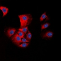
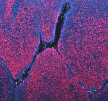
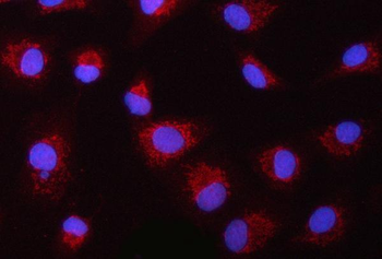
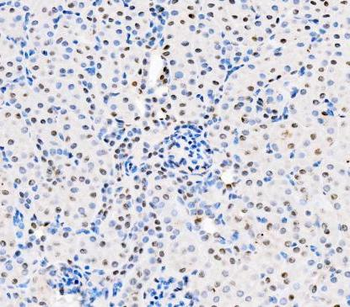
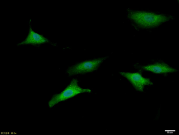
Immunofluorescent analysis of ASC staining in HepG2 cells. Formalin-fixed cells were permeabilized with 0.1% Triton X-100 in TBS for 5-10 minutes and blocked with 3% BSA-PBS for 30 minutes at room temperature. Cells were probed with the primary antibody in 3% BSA-PBS and incubated overnight at 4 °C in a hidified chamber. Cells were washed with PBST and incubated with a DyLight 594-conjugated secondary antibody (red) in PBS at room temperature in the dark. DAPI was used to stain the cell nuclei (blue).
Quick Database Links
UniProt Details
− No UniProt data available
NCBI Gene Details
− No NCBI Gene data available
Documents Download
Datasheet
Product Information
Request a Document
Protocol Information
WB
Western Blot (IB, immunoblot)
IHC
Immunohistochemistry
IF
Immunofluorescence
Filter by Applications
Filter by Species
Jun Sun 1, Qiuhua Zeng 2, Zhimin Wu 3, Lixin Huang 3, Tao Sun 3, Cong Ling 3, Baoyu Zhang 3, Chuan Chen 4, Hui Wang Berberine inhibits NLRP3 inflammasome activation and proinflammatory macrophage M1 polarization to accelerate peripheral nerve regeneration Neurotherapeutics, 2, e00347 (2024)
Applications
WB
Reactivity
Mouse
Xiaolan Huang 1, Xiangmin Luo 2, Suzhen Huang 2, Xiaoqing Chen 3, Lingling Qiu 2 Inhibition of FoxO1 alleviates polycystic ovarian syndrome by reducing inflammation and the immune response Funct Integr Genomics, (2024)
Applications
WB
Reactivity
Rat
Jia Shen 1, Xiaojun Ma Inhibition of the Foxo3/Txnip Axis Alleviates Ventilator-Induced Diaphragmatic Dysfunction by Downregulating MuRF1 Appl Biochem Biotechnol, (2025)
Applications
WB
Reactivity
Mouse
Sebastian Vogel et al. NLRP3 inflammasome-mediated platelet hyperreactivity in sickle cell mice is targetable by BTK inhibition Biochemical and Biophysical Research Communications,
Applications
IF
Reactivity
Mouse
ASC Rabbit Polyclonal Antibody (orb338943)

- 5.0
Based on 2 reviews
Participating in our Biorbyt product reviews program enables you to support fellow scientists by sharing your firsthand experience with our products.
Login to Submit a ReviewFilter by Rating
- 5 stars

- 4 stars
- 3 stars
- 2 stars
- 1 stars
Filter by Applications
Filter by Species
- 5 stars
The Rabbit Polyclonal ASC Antibody offers high antigen-binding affinity and effective signal amplification, supporting versatile detection requirements across various experimental platforms. Validated for IF, IHC, and WB applications, it facilitates the analysis of ASC protein expression and subcellular distribution. The antibody shows strong reproducibility and significant utility, making it well-suited for basic research focused on inflammatory responses, immune regulation, and cell death mechanisms.
- 5 stars
该ASC抗体为兔源多克隆抗体,具有良好的物种兼容性,可识别人、小鼠等多种来源样本中的ASC蛋白。产品支持免疫荧光(IF)、免疫组织化学(IHC)及Western blot(WB)等多种实验应用,适用于ASC蛋白表达及定位分析研究。作为炎症小体关键衔接蛋白的检测工具,该抗体具有较好的适用性和实验兼容性,能够满足组织、细胞及蛋白水平检测的需求。
Available Sizes
Select a size below
Choose Conjugation or Carrier Free Version
Free Secondary Antibody (20 ul)0/0
Please add an antibody product to your cart first.