You have no items in your shopping cart.
Description
Research Area
Neuroscience
Images & Validation
−Item 1 of 7
| Tested Applications | ELISA, ICC, IHC, WB |
|---|---|
| Dilution Range | WB (1:500) |
| Reactivity | Human, Mouse |
| Application Notes |
Key Properties
−| Antibody Type | Recombinant Antibody |
|---|---|
| Host | Rabbit |
| Clonality | Recombinant |
| Isotype | IgG |
| Clone No. | J18 |
| Immunogen | Human alpha synuclein AA 124-134: AYEMP-pS-EEGYQ-Cys |
| Target | Alpha Synuclein pSer129 |
| Purification | Affinity Purified |
| Conjugation | FITC |
Storage & Handling
−| Storage | Conjugated antibodies should be stored according to the product label |
|---|---|
| Buffer/Preservatives | 640.91mM DMSO, 136.36 mM Ethanolamine, 126.89 mM chlorides, 9.09mM phosphates, 9.09mM NaHCO3 |
| Concentration | 1 mg/ml |
| Expiration Date | 12 months from date of receipt. |
| Disclaimer | For research use only |
Alternative Names
−Alpha Synuclein, α-Synuclein, SNCA, alphaSYN, NACP, Non-A beta component of AD amyloid, Non-A4 component of amyloid precursor, Phosphorylated alpha synuclein, Phospho-alpha Synuclein (S129), Alpha-synuclein (phospho S129), Alpha Synuclein (phospho Ser129), Alpha-Synuclein Phospho Ser129, phospho-α-Synuclein (Ser129), Alpha Synuclein phospho Serine 129, Alpha Synuclein phospho Ser 129, Alpha Synuclein pSerine 129, Alpha Synuclein pSer 129, Alpha Synuclein phosphoSer 129, isoform NACP140, PARK1, PARK 1, PARK4, PARK 4, Parkinson disease (autosomal dominant, Lewy body) 4, Parkinson disease familial 1, SYN, Synuclein alpha, Synuclein alpha 140, Synuclein, alpha (non A4 component of amyloid precursor), SYUA_HUMAN
Similar Products
−- Item 1 of 5
Alpha Synuclein (pSer129) Antibody (FITC) [orb414137]
ELISA, ICC, IF, IHC, WB
Human, Mouse, Rat
Rabbit
Polyclonal
FITC
100 μl
Quality Guarantee
Explore bioreagents carefree to elevate your research. All our products are rigorously tested for performance. If a product does not perform as described on its datasheet, our scientific support team will provide expert troubleshooting, a prompt replacement, or a refund. For full details, please see our Terms & Conditions and Buying Guide. Contact us at [email protected].

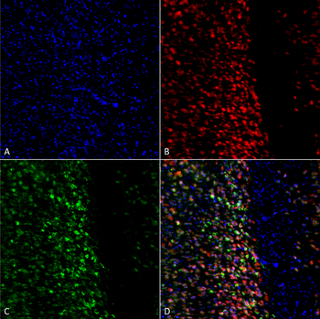
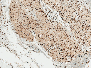
Immunohistochemistry analysis using Rabbit Anti-Alpha Synuclein pSer129 Monoclonal Antibody, Clone J18. Tissue: Brain. Species: Mouse. Primary Antibody: Rabbit Anti-Alpha Synuclein pSer129 Monoclonal Antibody at 1:10000. Secondary Antibody: anti-rabbit HRP. C57/BL6 mice were injected with 5 ug sonicated mouse recombinant alpha synuclein PFFs at 8 weeks of age. Mice were unilaterally injected in the dorsal striatum (bregma AP + 0.2 mm, L +/1 2.0 mm, V - 3.0 mm) and sacrificed 30 days post-injection. (A) contralateral cortex. (B) ipsilateral cortex. (C) contralateral striatum. (D) ipsilateral striatum.
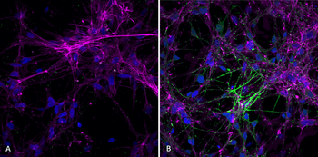
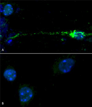
Immunocytochemistry/Immunofluorescence analysis using Rabbit Anti-Alpha Synuclein (pSer129) Monoclonal Antibody, Clone J18. Tissue: iPSC-derived neurons. Species: Human. Primary Antibody: Rabbit Anti-Alpha Synuclein (pSer129) Monoclonal Antibody at 1:1000 for O/N at 4°C. Secondary Antibody: Anti-Rabbit: A488 at 1:1000 for 1 hour at RT. Magnification: 40X. Nuclear stain: Hoechst- 20 min, RT (blue). Actin stain: Phalloidin-647- 20 min, RT (magenta). 4K cells per well. A) negative control; no fibrils added to well. B) 7 days after addition of active recombinant human pre-formed fibrils (Type 1).
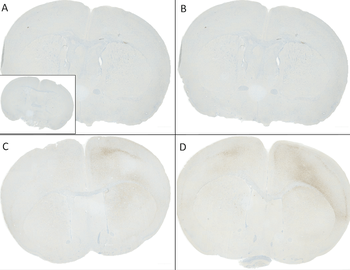
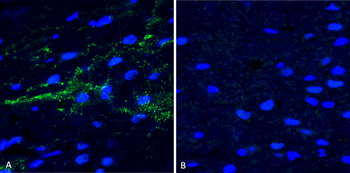
Immunohistochemistry analysis using Rabbit Anti-Alpha Synuclein pSer129 Monoclonal Antibody, Clone J18. Tissue: Brain. Species: Mouse. Primary Antibody: Rabbit Anti-Alpha Synuclein pSer129 Monoclonal Antibody at 1:10000. Secondary Antibody: anti-rabbit HRP. C57/BL6 mice were injected with sonicated recombinant mouse alpha synuclein monomers or fibrils at 8 weeks of age. Mice were unilaterally injected in the dorsal striatum (bregma AP + 0.2 mm, L +/1 2.0 mm, V - 3.0 mm) and sacrificed 30 days post-injection. (A) 1.25 uL mouse alpha synuclein monomers. (B) 2.5 uL mouse alpha synuclein monomers. (C) 2.5 ug alpha synuclein PFFs. (D) 5 ug alpha synuclein PFFs. Inset: PBS (negative control).
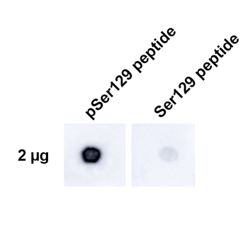
Dot Blot analysis using Rabbit Anti-Alpha Synuclein pSer129 Monoclonal Antibody, Clone J18. Tissue: alpha synuclein peptide. Primary Antibody: Rabbit Anti-Alpha Synuclein pSer129 Monoclonal Antibody at 1:500 for 2 hours at RT with shaking. Secondary Antibody: Goat anti-rabbit IgG:HRP at 1:4000 for 1 hour at RT with shaking. Phospho peptide sequence: AYEMP-pS-EEGYQ. Non-phospho peptide sequence: AYEMPSEEGYQ. This sequence is the same for human, mouse, and rat.
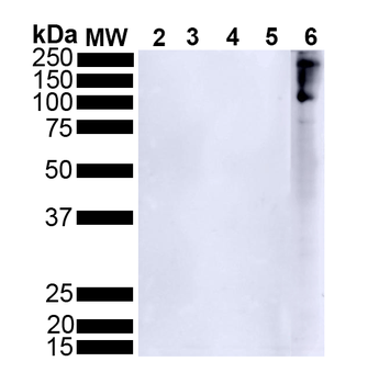
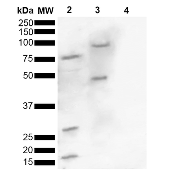
Western Blot analysis of Human Alpha Synuclein showing detection of Alpha Synuclein pSer129 protein using Rabbit Anti-Alpha Synuclein pSer129 Monoclonal Antibody, Clone J18. Lane 1: MW ladder. Lane 2: 0.5 ug human alpha synuclein monomer. Lane 3: 2 ug human alpha synuclein monomer. Lane 4: 0.5 ug human alpha synuclein PFFs. Lane 5: 2 ug human alpha synuclein PFFs. Lane 6: 15 ug human Parkinson's Disease brain lysate. Block: 5% BSA in TBST. Primary Antibody: Rabbit Anti-Alpha Synuclein pSer129 Monoclonal Antibody at 1:500 for 2 hours at RT with shaking. Secondary Antibody: Goat anti-rabbit IgG:HRP at 1:4000 for 1 hour at RT with shaking. Color Development: Chemiluminescent for HRP (Moss) for 5 min in RT.
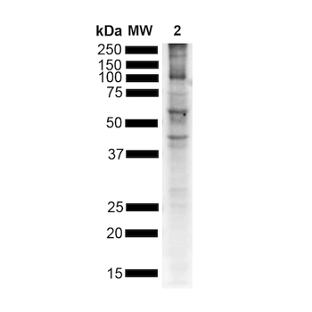
Western Blot analysis of Mouse Brain showing detection of Alpha Synuclein pSer129 protein using Rabbit Anti-Alpha Synuclein pSer129 Monoclonal Antibody, Clone J18. Lane 1: MW ladder. Lane 2: Mouse brain. Load: 15 ug. Block: 5% BSA in TBST. Primary Antibody: Rabbit Anti-Alpha Synuclein pSer129 Monoclonal Antibody at 1:500 for 2 hours at RT with shaking. Secondary Antibody: Goat anti-rabbit IgG:HRP at 1:4000 for 1 hour at RT with shaking. Color Development: Chemiluminescent for HRP (Moss) for 5 min in RT.
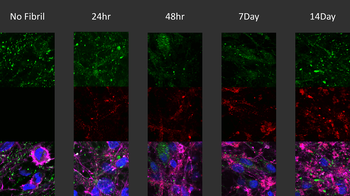
Immunocytochemistry / Immunofluorescence analysis of human iPSC-derived neurons treated with 2.5μg ATTO 594 labeled type I alpha-synuclein pre-formed fibrils for up to 14 days. Cells seeded at 8k cells per well. Green: mouse anti-alpha synuclein (pSer129) monoclonal antibody 1:5000; Red: alpha-synuclein PFFs; Pink: actin; Blue: Hoechst / DNA.
Quick Database Links
UniProt Details
− No UniProt data available
NCBI Gene Details
− No NCBI Gene data available
NCBI Reference Sequences
−Associated Accession Numbers
Curated reference sequences for the gene transcript and protein product| Protein | NP_000336.1 |
|---|
Documents Download
Datasheet
Product Information
Request a Document
Protocol Information
WB
Western Blot (IB, immunoblot)
IHC
Immunohistochemistry
ICC
Immunocytochemistry
ELISA
Enzyme-linked Immunosorbent Assay (EIA)
Alpha Synuclein (pSer129) Antibody (FITC) (orb612726)

- 0.0
Based on 0 reviews
Participating in our Biorbyt product reviews program enables you to support fellow scientists by sharing your firsthand experience with our products.
Login to Submit a ReviewAvailable Sizes
Select a size below
Choose Conjugation or Carrier Free Version
Free Secondary Antibody (20 ul)0/0
Please add an antibody product to your cart first.