You have no items in your shopping cart.
Featured
Description
Research Area
Neuroscience
Images & Validation
−Item 1 of 5
| Tested Applications | DOT, ELISA, ICC, IF, WB |
|---|---|
| Dilution Range | WB (1:1000); DB (1:1000); ICC/IF (1:100); ELISA (1:1000) |
| Reactivity | Human, Mouse, Rat |
| Application Notes |
Key Properties
−| Host | Mouse |
|---|---|
| Clonality | Monoclonal |
| Isotype | IgG1 |
| Clone No. | 3C11 |
| Immunogen | Human alpha synuclein monomer |
| Target | Alpha Synuclein |
| Molecular Weight | 30 kDa |
| Purification | Protein G Purified |
| Conjugation | Unconjugated |
Storage & Handling
−| Storage | Maintain refrigerated at 2-8°C for up to 2 weeks. For long term storage store at -20°C in small aliquots to prevent freeze-thaw cycles. |
|---|---|
| Buffer/Preservatives | PBS pH 7.4, 50% glycerol, 0.09% sodium azide. Storage buffer changes when conjugated. |
| Concentration | 1 mg/ml |
| Expiration Date | 12 months from date of receipt. |
| Disclaimer | For research use only |
Alternative Names
−Alpha Synuclein, α-Synuclein, SNCA, Snca, SYN, alphaSYN, NACP, Non-A beta component of AD amyloid, Non-A4 component of amyloid precursor, Parkinson disease familial 1, PARK1, PARK 1, PARK4, PARK 4, Parkinson disease (autosomal dominant, Lewy body) 4
Similar Products
−- Item 1 of 15
Alpha Synuclein Recombinant Rabbit Monoclonal Antibody [orb1227214]
FC, ICC, IF, IHC-Fr, IHC-P, WB
Mouse, Rat
Human, Rat
Rabbit
Recombinant
Unconjugated
50 μl, 100 μl, 25 μl - Item 1 of 7
Alpha-Synuclein Rabbit Polyclonal Antibody [orb11442]
WB
Human, Mouse, Rat
Human, Mouse, Rat
Rabbit
Polyclonal
Unconjugated
50 μl, 100 μl - Item 1 of 7
Alpha Synuclein (pSer129) Antibody (RPE) [orb612730]
ELISA, ICC, IHC, WB
Human, Mouse
Rabbit
Recombinant
RPE
100 μg - Item 1 of 7
Alpha Synuclein (pSer129) Antibody (APC) [orb612723]
ELISA, ICC, IHC, WB
Human, Mouse
Rabbit
Recombinant
APC
100 μg - Item 1 of 7
Alpha Synuclein (pSer129) Antibody (HRP) [orb612727]
ELISA, ICC, IHC, WB
Human, Mouse
Rabbit
Recombinant
HRP
100 μg


Quality Guarantee
Explore bioreagents carefree to elevate your research. All our products are rigorously tested for performance. If a product does not perform as described on its datasheet, our scientific support team will provide expert troubleshooting, a prompt replacement, or a refund. For full details, please see our Terms & Conditions and Buying Guide. Contact us at [email protected].
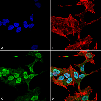
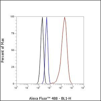
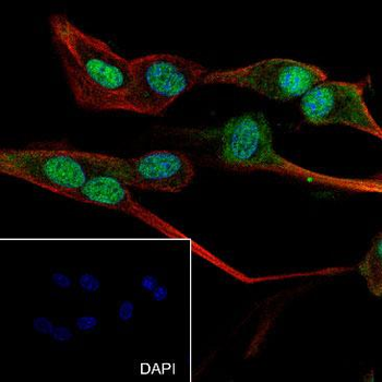
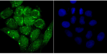
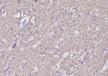
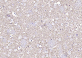
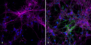
Immunocytochemistry/Immunofluorescence analysis using Mouse Anti-Alpha Synuclein Monoclonal Antibody, Clone 3C11. Tissue: Neuroblastoma cell line (SK-N-BE). Species: Human. Fixation: 4% Formaldehyde for 15 min at RT. Primary Antibody: Mouse Anti-Alpha Synuclein Monoclonal Antibody at 1:100 for 60 min at RT. Secondary Antibody: Goat Anti-Mouse ATTO 488 at 1:200 for 60 min at RT. Counterstain: Phalloidin Texas Red F-Actin stain; DAPI (blue) nuclear stain at 1:1000, 1:5000 for 60 min at RT, 5 min at RT. Localization: Cytoplasm: weak; Nucleus: Med. Magnification: 60X. (A) DAPI (blue) nuclear stain. (B) Phalloidin Texas Red F-Actin stain. (C) Alpha Synuclein Antibody. (D) Composite.
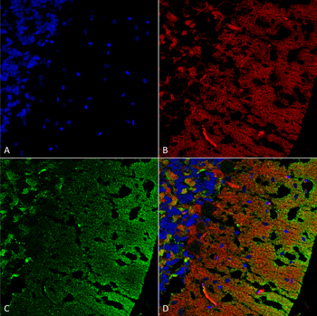
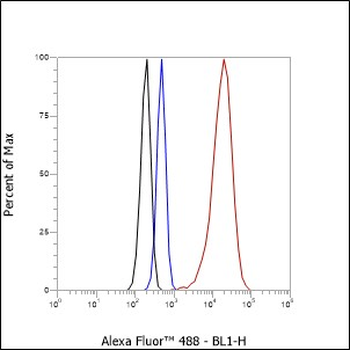
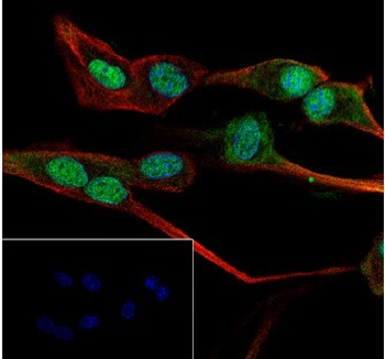
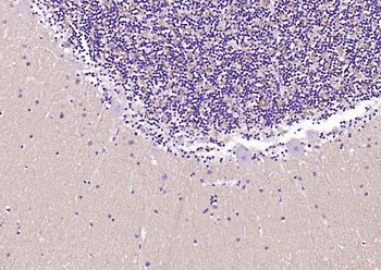
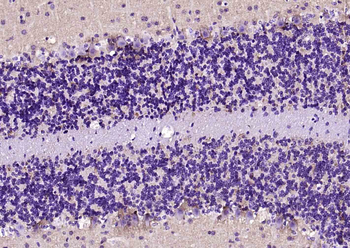
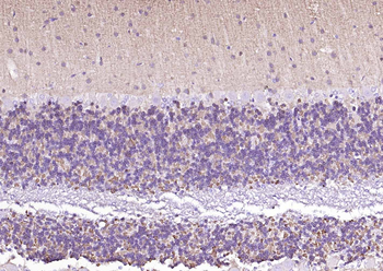
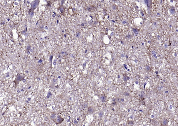
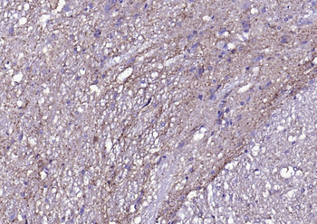
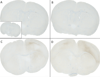
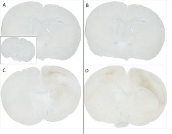
Immunohistochemistry analysis using Mouse Anti-Alpha Synuclein Monoclonal Antibody, Clone 3C11. Tissue: cerebellum. Species: Rat. Fixation: Formalin fixed, paraffin embedded. Primary Antibody: Mouse Anti-Alpha Synuclein Monoclonal Antibody at 1:25 for 1 hour at RT. Secondary Antibody: Goat Anti-Mouse IgG: Alexa Fluor 488. Counterstain: Actin-binding Phalloidin-Alexa Fluor 633; DAPI (blue) nuclear stain. Magnification: 63X. (A) DAPI (blue) nuclear stain. (B) Phalloidin Alexa Fluor 633 F-Actin stain. (C) Alpha Synuclein Antibody (D) Composite.
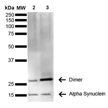
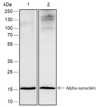
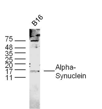
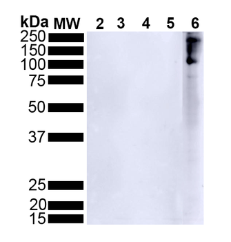
Western Blot analysis of Human Brain showing detection of 14 kDa Alpha Synuclein protein using Mouse Anti-Alpha Synuclein Monoclonal Antibody, Clone 3C11. Lane 1: Molecular Weight Ladder (MW). Lane 2: Parkinson brain cell lystate. Lane 3: Human brain cell lysate. Load: 15 μg. Block: 5% Skim Milk in 1X TBST. Primary Antibody: Mouse Anti-Alpha Synuclein Monoclonal Antibody at 1:1000 for 2 hours at RT. Secondary Antibody: Goat Anti-Mouse HRP:IgG at 1:3000 for 1 hour at RT. Color Development: ECL solution (Super Signal West Pico) for 5 min in RT. Predicted/Observed Size: 14 kDa. Other Band (s): 100 kDa (oligomer).
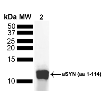
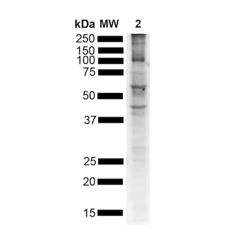
Western Blot analysis of Human Truncated Alpha Synuclein Protein showing detection of Alpha Synuclein protein using Mouse Anti-Alpha Synuclein Monoclonal Antibody, Clone 3C11. Lane 1: MW Ladder. Lane 2: hASYN aa 1-114 (5 uL). Load: 5 uL. Block: 5% Skim Milk Powder in TBST. Primary Antibody: Mouse Anti-Alpha Synuclein Monoclonal Antibody at 1:1000 for 2 hours at RT with shaking. Secondary Antibody: Goat anti-mouse IgG:HRP at 1:4000 for 1 hour at RT with shaking. Color Development: Chemiluminescent for HRP (Moss) for 5 min in RT.
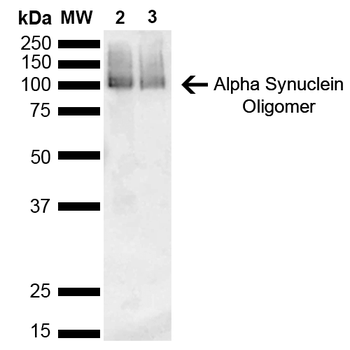
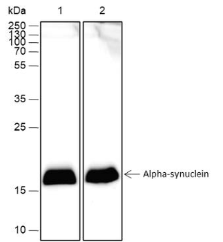
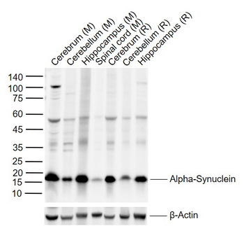
Western Blot analysis of Mouse, Rat Brain showing detection of 14 kDa Alpha Synuclein protein using Mouse Anti-Alpha Synuclein Monoclonal Antibody, Clone 3C11. Lane 1: Molecular Weight Ladder (MW). Lane 2: Mouse brain cell lysate. Lane 3: Rat brain cell lysate. Load: 15 μg. Block: 5% Skim Milk in 1X TBST. Primary Antibody: Mouse Anti-Alpha Synuclein Monoclonal Antibody at 1:1000 for 2 hours at RT. Secondary Antibody: Goat Anti-Mouse HRP:IgG at 1:3000 for 1 hour at RT. Color Development: ECL solution (Super Signal West Pico) for 5 min in RT. Predicted/Observed Size: 14 kDa. Other Band (s): ~30 kDa (dimer).
Quick Database Links
UniProt Details
− No UniProt data available
NCBI Gene Details
− No NCBI Gene data available
NCBI Reference Sequences
−Associated Accession Numbers
Curated reference sequences for the gene transcript and protein product| Protein | NP_000336.1 |
|---|
Documents Download
Datasheet
Product Information
Request a Document
Protocol Information
WB
Western Blot (IB, immunoblot)
IF
Immunofluorescence
ICC
Immunocytochemistry
ELISA
Enzyme-linked Immunosorbent Assay (EIA)
DOT
Dot Blot
Alpha Synuclein Antibody (orb413297)

- 0.0
Based on 0 reviews
Participating in our Biorbyt product reviews program enables you to support fellow scientists by sharing your firsthand experience with our products.
Login to Submit a ReviewAvailable Sizes
Select a size below
Choose Conjugation or Carrier Free Version
Free Secondary Antibody (20 ul)0/0
Please add an antibody product to your cart first.