You have no items in your shopping cart.
Description
Research Area
Metabolism Research
Images & Validation
−Item 1 of 4
| Tested Applications | FC, IF, IHC-P, WB |
|---|---|
| Dilution Range | IF - 1:100, WB - 1:1000, IHC-P - 1:50-100, FC - 1:10-50 |
| Reactivity | Human |
Key Properties
−| Host | Rabbit |
|---|---|
| Clonality | Polyclonal |
| Isotype | Rabbit IgG |
| Immunogen | This ALDH1A1 antibody is generated from rabbits immunized with human ALDH1A1 recombinant protein. |
| Target | ALDH1A1 (HGNC:402) |
| Molecular Weight | 54862 Da |
| Conjugation | Unconjugated |
Storage & Handling
−| Storage | Maintain refrigerated at 2-8°C for up to 2 weeks. For long term storage store at -20°C in small aliquots to prevent freeze-thaw cycles |
|---|---|
| Form/Appearance | Purified polyclonal antibody supplied in PBS with 0.09% (W/V) sodium azide. This antibody is purified through a protein A column, followed by peptide affinity purification. |
| Expiration Date | 12 months from date of receipt. |
| Disclaimer | For research use only |
Alternative Names
−Retinal dehydrogenase 1, RALDH 1, RalDH1, ALDH-E1, ALHDII, Aldehyde dehydrogenase family 1 member A1, Aldehyde dehydrogenase, cytosolic, ALDH1A1, ALDC, ALDH1, PUMB1
Similar Products
−- Item 1 of 10
Retinal dehydrogenase 1 ALDH1A1 Rabbit Polyclonal Antibody [orb570330]
ELISA, FC, ICC, IF, IHC, WB
Human, Mouse, Rat
Rabbit
Polyclonal
Unconjugated
100 μg - Item 1 of 5
ALDH1A1 Rabbit Polyclonal Antibody [orb155630]
IF, IHC-Fr, IHC-P, WB
Bovine, Equine, Porcine, Rabbit, Rat, Sheep
Human, Mouse
Rabbit
Polyclonal
Unconjugated
50 μl, 100 μl, 200 μl - Item 1 of 7
ALDH1A1 Recombinant Rabbit Monoclonal Antibody [orb1152015]
IF, IHC-Fr, IHC-P, WB
Mouse
Human, Mouse
Rabbit
Recombinant
Unconjugated
50 μl, 100 μl, 25 μl - Item 1 of 5
ALDH1A1 Antibody (Center) [orb1935603]
FC, IF, IHC-P, WB
Mouse
Human
Rabbit
Polyclonal
Unconjugated
400 μl - Item 1 of 4
ALDH1A1 rabbit pAb Antibody [orb766762]
ELISA, IF, IHC, WB
Human, Mouse, Rat
Polyclonal
Unconjugated
50 μl, 100 μl
Quality Guarantee
Explore bioreagents carefree to elevate your research. All our products are rigorously tested for performance. If a product does not perform as described on its datasheet, our scientific support team will provide expert troubleshooting, a prompt replacement, or a refund. For full details, please see our Terms & Conditions and Buying Guide. Contact us at [email protected].
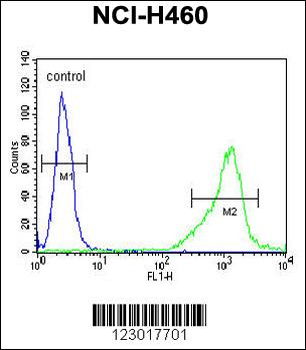
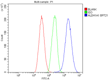
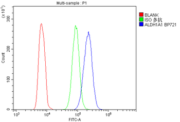
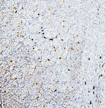
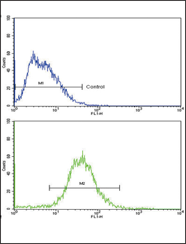
ALDH1A1 Antibody flow cytometric analysis of NCI-H460 cells (right histogram) compared to a negative control cell (left histogram). FITC-conjugated goat-anti-rabbit secondary antibodies were used for the analysis.
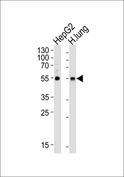
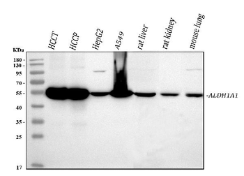
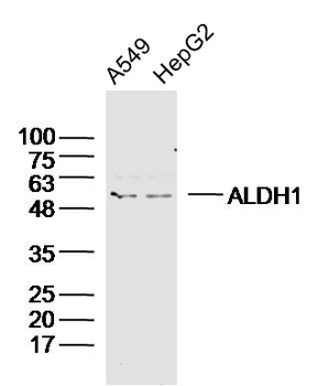
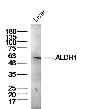
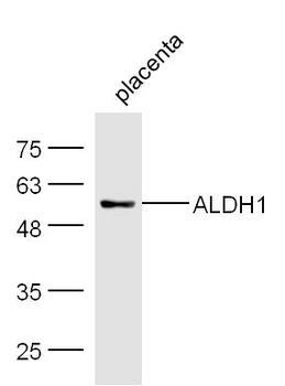
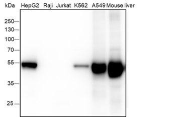
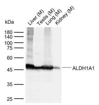
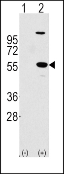
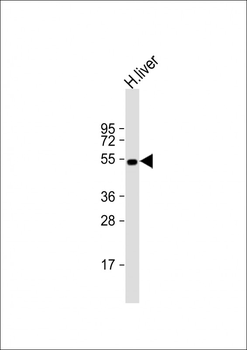
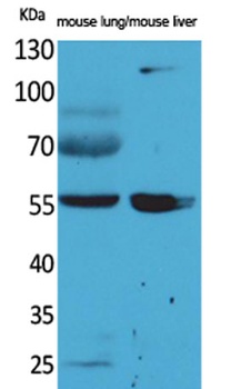
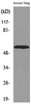
Western blot analysis of lysates from HepG2 cell line and human lung tissue lysate (from left to right), using ALDH1A1 Antibody. Diluted at 1:1000 at each lane. A goat anti-rabbit IgG H&L (HRP) at 1:5000 dilution was used as the secondary antibody. Lysates at 35ug per lane.
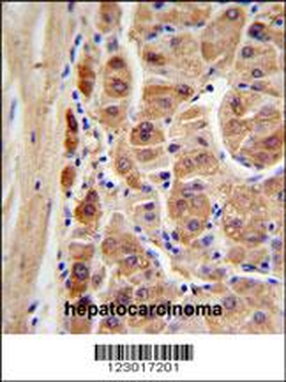
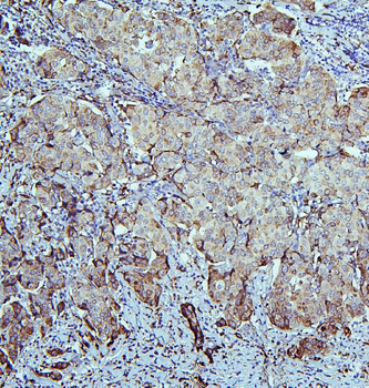
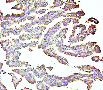
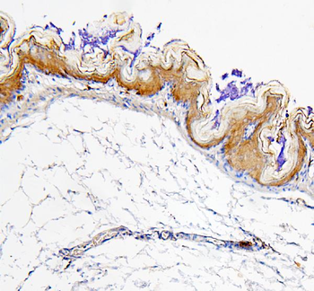
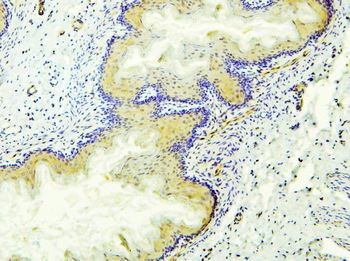
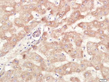
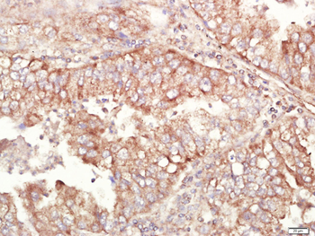
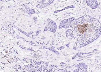
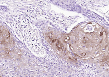
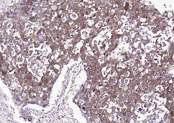
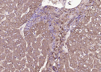
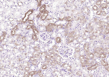
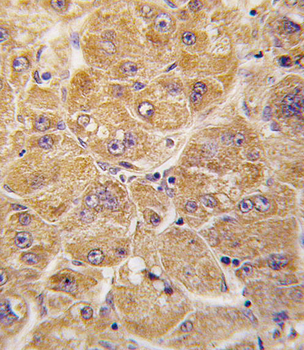
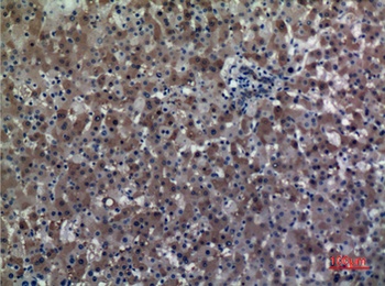
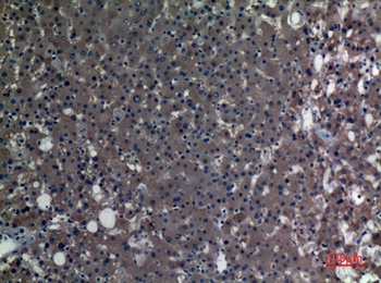
ALDH1A1 Antibody IHC analysis in formalin fixed and paraffin embedded human hepatocarcinoma followed by peroxidase conjugation of the secondary antibody and DAB staining. This data demonstrates the use of the ALDH1A1 Antibody for immunohistochemistry. Clinical relevance has not been evaluated.
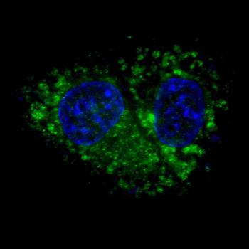
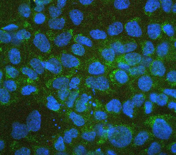
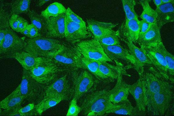
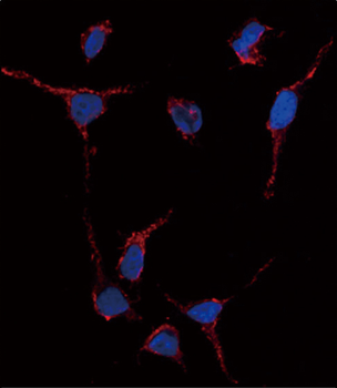
Fluorescent confocal image of HepG2 cells stained with ALDH1A1 antibody. HepG2 cells were fixed with 4% PFA (20 min), permeabilized with Triton X-100 (0.2%, 30 min). Cells were then incubated with ALDH1A1 primary antibody (1:100, 2 h at room temperature). For secondary antibody, Alexa Fluor 488 conjugated donkey anti-rabbit antibody (green) was used (1:1000, 1 h). Nuclei were counterstained with Hoechst 33342 (blue) (10 μg/ml, 5 min). ALDH1A1 immunoreactivity is localized to the cytoplasm of HepG2 cells.
Quick Database Links
UniProt Details
− No UniProt data available
NCBI Reference Sequences
−Associated Accession Numbers
Curated reference sequences for the gene transcript and protein product| Protein | NP_000680.2 |
|---|
Documents Download
Datasheet
Product Information
Request a Document
Protocol Information
WB
Western Blot (IB, immunoblot)
IHC-P
Immunohistochemistry Paraffin
FC
Flow Cytometry
IF
Immunofluorescence
ALDH1A1 Antibody (orb1935602)

- 0.0
Based on 0 reviews
Participating in our Biorbyt product reviews program enables you to support fellow scientists by sharing your firsthand experience with our products.
Login to Submit a ReviewAvailable Sizes
Select a size below
Choose Conjugation or Carrier Free Version
Free Secondary Antibody (20 ul)0/0
Please add an antibody product to your cart first.