You have no items in your shopping cart.
Description
Research Area
Metabolism Research
Images & Validation
−Item 1 of 5
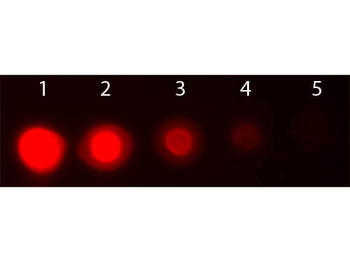
| Tested Applications | DOT, ELISA |
|---|---|
| Dilution Range | ELISA: User Optimized, FC: User Optimized, IHC: User Optimized, IF: User Optimized, WB: User Optimized |
| Reactivity | Human, Mouse, Rat |
| Application Notes |
Key Properties
−| Antibody Type | Primary Antibody |
|---|---|
| Host | Mouse |
| Clonality | Monoclonal |
| Isotype | IgG1 |
| Clone No. | 25F6.F6.D8 |
| Immunogen | Anti-AKT3 Antibody was prepared from tissue culture supernatant by Protein A affinity chromatography using a synthetic peptide corresponding to internal residues of human AKT3 protein. |
| Target | AKT3 |
| Purity | Anti-AKT3 antibody is directed against human AKT3. The antibody detects both unphosphorylated and phosphorylated forms of the protein. Anti-AKT3 antibody was purified from tissue culture by Protein A chromatography. Cross reactivity with AKT3 from other species has not been determined, however, the sequence of the immunogen shows 100% identity to human, mouse, and rat, therefore, cross reactivity is expected. Cross-reactivity with AKT2 and AKT has not been determined. |
| Conjugation | RPE |
Storage & Handling
−| Storage | Store vial at 4° C prior to restoration. Restore with deionized water (or equivalent). This product is stable at 4° C as an undiluted liquid. Dilute only prior to immediate use. Centrifuge product if not completely clear after standing at room temperature. Do not freeze after reconstitution. Store reagent in the dark. Use subdued lighting during handling and incubation of cells prior to analysis. |
|---|---|
| Form/Appearance | Lyophilized |
| Buffer/Preservatives | Preservative: None. Stabilizer: 10 mg/mL Bovine Serum Albumin (rAlbumin) - Immunoglobulin and Protease free; Buffer: 0.02 M Potassium Phosphate, 0.5 M Sodium Chloride, pH 7.2 |
| Concentration | 1 mg/mL |
| Expiration Date | 12 months from date of receipt. |
| Disclaimer | For research use only |
Alternative Names
−Mouse anti-AKT3 antibody PE conjugation, phycoerythrin conjugated Mouse anti-AKT 3 antibody, AKT-3, PKB antibody, PKB gamma antibody, PKBGAMMA antibody, PRKBG antibody, Protein kinase Akt 3 antibody, Protein kinase B gamma antibody, RAC-gamma serine/threonine-protein kinase, RAC-PK-gamma
Similar Products
−- Item 1 of 2
Quality Guarantee
Explore bioreagents carefree to elevate your research. All our products are rigorously tested for performance. If a product does not perform as described on its datasheet, our scientific support team will provide expert troubleshooting, a prompt replacement, or a refund. For full details, please see our Terms & Conditions and Buying Guide. Contact us at [email protected].
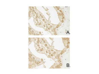
Immunohistochemistry of Mouse Anti-AKT3 antibody. Tissue: human prostate carcinoma. A) AKT-3 antibody produced using CELLine, B) AKT-3 antibody produced using roller bottle. Fixation: formalin fixed paraffin embedded. Antigen retrieval: not required. Primary antibody: AKT-3 antibody at 10 µg/ml for 1 h at RT. Secondary antibody: Peroxidase mouse secondary antibody at 1:10000 for 1 h at RT. Localization: AKT3 is nuclear and occasionally cytoplasmic. Staining: AKT3 as precipitated brown signal with hematoxylin purple nuclear counterstain.
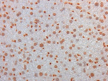
Immunohistochemistry of Mouse Monoclonal anti AKT3 Antibody in Mouse Embryonic Kidney. Tissue: Mouse Liver. Fixation: FFPE buffered formalin 10% conc. Ag Retrieval: Heat, Citrate pH6.2. Pressure Cooker. Primary antibody: anti-AKT3 at 2 ug/ml for 1.5 hour @ room Temp. Secondary Ab: MOUSE ON MOUSE HRP POLYMER 45" RT.
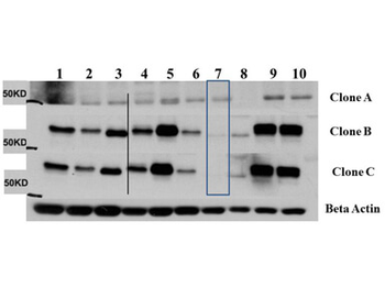
Western Blot of Mouse Anti-AKT3 antibody. Lane 1: C2C12. Lane 2: MEF#1. Lane 3: MEF#2. Lane 4: A549. Lane 5: Calu-1. Lane 6: PC3. Lane 7: HepG2. Lane 8: Jurkat. Lane 9: SKOV3. Lane 10: 293T. Load: 35 µg per lane. Primary antibody: AKT-3 antibody at 1:1000 for overnight at 4°C. Secondary antibody: Anti mouse secondary antibody at 1:20000 for 1 h at RT. Block: 5% BLOTTO overnight at 4°C. Predicted/Observed size: 56 kDa for AKT3.
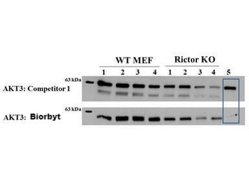
Western Blot of Mouse anti-AKT3 antibody. Lane 1: Control. Lane 2: Rapa. Lane 3: T50. Lane 4: T250. Lane 5: Control. Lane 6: Rapa. Lane 7: T50. Lane 8: T250. Lane 9: AKT3 null. Load: 35 µg per lane. Primary antibody: AKT-3 antibody at 1:1000 for overnight at 4°C. Secondary antibody: Anti mouse secondary antibody at 1:20000 for 1 h at RT. Block: 5% BLOTTO overnight at 4°C. Predicted/Observed size: 56 kDa for AKT3.
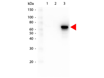
Western Blot of Mouse anti-AKT3 antibody. Lane 1: GST Tagged recombinant AKT1. Lane 2: GST Tagged recombinant AKT2. Lane 3: GST Tagged recombinant AKT3. Load: 25 ng per lane. Primary antibody: AKT3 antibody at 1:1000 for overnight at 4°C. Secondary antibody: Peroxidase mouse secondary antibody at 1:40000 for 30 min at RT. Block: orb348637 for 30 min at RT. Predicted/Observed size: 78 kDa for AKT3. Other band(s): none.
Quick Database Links
UniProt Details
− No UniProt data available
NCBI Reference Sequences
−Associated Accession Numbers
Curated reference sequences for the gene transcript and protein product| Protein | NP_001193658.1 |
|---|
Documents Download
Datasheet
Product Information
Request a Document
Protocol Information
ELISA
Enzyme-linked Immunosorbent Assay (EIA)
DOT
Dot Blot
AKT3 Antibody (RPE) (orb344550)

- 0.0
Based on 0 reviews
Participating in our Biorbyt product reviews program enables you to support fellow scientists by sharing your firsthand experience with our products.
Login to Submit a ReviewAvailable Sizes
Select a size below
Free Secondary Antibody (20 ul)0/0
Please add an antibody product to your cart first.