You have no items in your shopping cart.
Featured
Description
Research Area
Signal Transduction
Images & Validation
−Item 1 of 9
| Tested Applications | ICC, IF, IHC-Fr, IHC-P, WB |
|---|---|
| Dilution Range | WB=1:500-2000, IHC-P=1:100-500, IHC-F=1:100-500, ICC/IF=1:100-500, IF=1:100-500 |
| Reactivity | Human, Mouse, Rat |
| Predicted Reactivity | Bovine, Canine, Gallus, Porcine, Rabbit, Sheep |
Related Conjugates & Formulations
−Key Properties
−| Antibody Type | Primary Antibody |
|---|---|
| Host | Mouse |
| Clonality | Polyclonal |
| Isotype | IgG |
| Immunogen | KLH conjugated synthetic peptide derived from human AKT-1 (401-479/479aa) |
| Target | AKT1 |
| Molecular Weight | 60 kDa |
| Purification | Affinity purified by Protein A |
| Conjugation | Unconjugated |
Storage & Handling
−| Storage | Maintain refrigerated at 2-8°C for up to 2 weeks. For long term storage store at -20°C in small aliquots to prevent freeze-thaw cycles. |
|---|---|
| Form/Appearance | Liquid |
| Buffer/Preservatives | 0.01M TBS (pH7.4) with 1% rAlbumin, 0.02% Proclin300 and 50% Glycerol. |
| Concentration | 1mg/ml |
| Expiration Date | 12 months from date of receipt. |
| Disclaimer | For research use only |
Alternative Names
−AKT; PKB; PKB-ALPHA; PRKBA; RAC; RAC-ALPHA; LTR-akt; PKB/Akt; PKBalpha; O57513_CHICK; akt1; RAC-PK-alpha; 2.7.11.1; AKT1_HUMAN; Protein kinase B (PKB); Protein kinase B alpha (PKB alpha); Proto-oncogene c-Akt; AKT1_MOUSE; AKT1 kinase; Thymoma viral proto-oncogene; AKT1_RAT;
Similar Products
−- Item 1 of 5
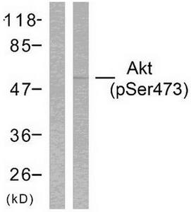
Akt (Phospho-Ser473) Antibody [orb127667]
IF, IHC, WB
Human, Mouse, Rat
Rabbit
Polyclonal
Unconjugated
50 μg, 25 μg, 100 μg, 200 μg - Item 1 of 2
Akt (Phospho-Ser473) Antibody [orb222951]
IHC, WB
Human, Mouse, Rat
Rabbit
Polyclonal
Unconjugated
50 μg, 25 μg, 100 μg, 200 μg - Item 1 of 2
Mouse Akt1 Antibody (N-term) [orb32910]
IHC-P, WB
Human
Mouse
Rabbit
Polyclonal
Unconjugated
50 μl, 100 μl - Item 1 of 3
AKT1,2,3/AKT1 Rabbit Polyclonal Antibody [orb1098048]
ELISA, FC, ICC, IF, WB
Human, Mouse, Rat
Rabbit
Polyclonal
Unconjugated
100 μg - Item 1 of 2
Akt (Phospho-Thr308) Antibody [orb222952]
IHC, WB
Human, Mouse, Rat
Rabbit
Polyclonal
Unconjugated
50 μg, 25 μg, 100 μg, 200 μg
_antibody_orb127667_wb_1.jpg)
_antibody_orb127667_wb_2.jpg)
_antibody_orb127667_ihc_p_1.jpg)
_antibody_orb127667_if_1.jpg)
Quality Guarantee
Explore bioreagents carefree to elevate your research. All our products are rigorously tested for performance. If a product does not perform as described on its datasheet, our scientific support team will provide expert troubleshooting, a prompt replacement, or a refund. For full details, please see our Terms & Conditions and Buying Guide. Contact us at [email protected].
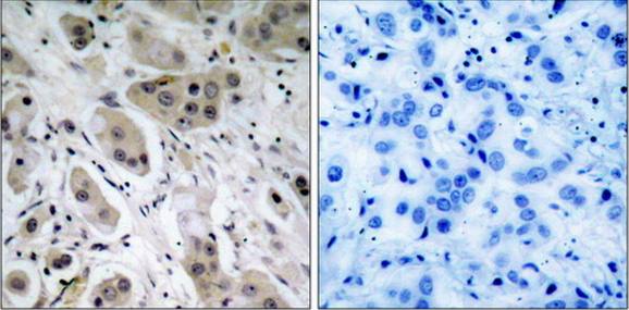
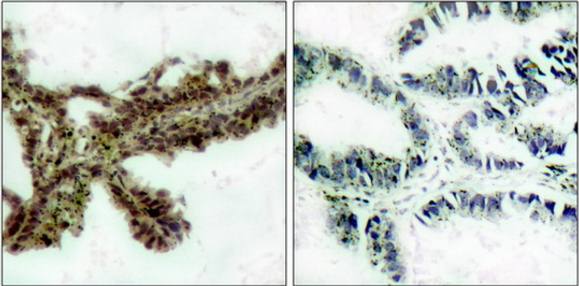
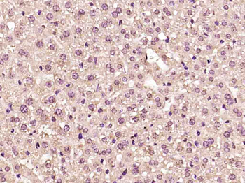
Paraformaldehyde-fixed, paraffin embedded (human memmery cancer), Antigen retrieval by boiling in sodium citrate buffer (pH6.0) for 15 min, Block endogenous peroxidase by 3% hydrogen peroxide for 20 minutes, Blocking buffer (normal goat serum) at 37°C for 30 min, Antibody incubation with (AKT1) Polyclonal Antibody, Unconjugated at 1:500 overnight at 4°C, followed by a conjugated secondary for 20 minutes and DAB staining.
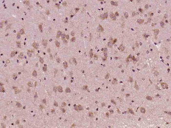
Paraformaldehyde-fixed, paraffin embedded (Mouse brain), Antigen retrieval by boiling in sodium citrate buffer (pH6.0) for 15 min, Block endogenous peroxidase by 3% hydrogen peroxide for 20 minutes, Blocking buffer (normal goat serum) at 37°C for 30 min, Antibody incubation with (AKT1) Monoclonal Antibody, Unconjugated (orb11276) at 1:400 overnight at 4°C, followed by operating according to SP Kit (Mouse) instructionsand DAB staining.
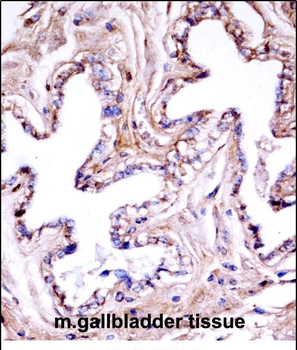
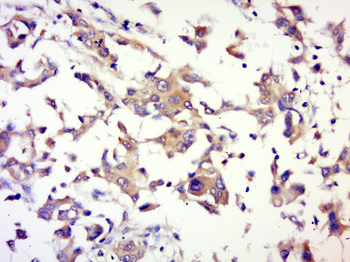
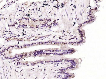
Paraformaldehyde-fixed, paraffin embedded (mouse liver), Antigen retrieval by boiling in sodium citrate buffer (pH6.0) for 15 min, Block endogenous peroxidase by 3% hydrogen peroxide for 20 minutes, Blocking buffer (normal goat serum) at 37°C for 30 min, Antibody incubation with (AKT1) Polyclonal Antibody, Unconjugated (orb11276) at 1:200 overnight at 4°C, followed by operating according to SP Kit (Rabbit) instructionsand DAB staining.
Paraformaldehyde-fixed, paraffin embedded (rat lung), Antigen retrieval by boiling in sodium citrate buffer (pH6.0) for 15 min, Block endogenous peroxidase by 3% hydrogen peroxide for 20 minutes, Blocking buffer (normal goat serum) at 37°C for 30 min, Antibody incubation with (AKT1) Monoclonal Antibody, Unconjugated (orb11276) at 1:200 overnight at 4°C, followed by operating according to SP Kit (Mouse) instructionsand DAB staining.
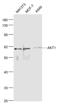
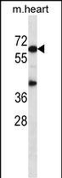
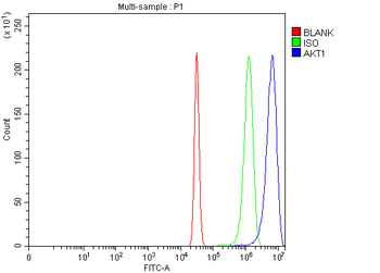
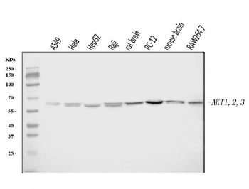
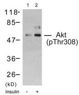
Sample: NIH/3T3 (Mouse) Cell Lysate at 30 ug, MCF-7 (Human) Cell Lysate at 30 ug, A549 (Human) Cell Lysate at 30 ug, Primary: Anti-AKT1 (orb11276) at 1/1000 dilution, Secondary: IRDye800CW Goat Anti-Mouse IgG at 1/20000 dilution, Predicted band size: 56 kD, Observed band size: 60 kD.
Tissue/Cell: Hela cell, 4% Paraformaldehyde-fixed, Triton X-100 at room temperature for 20 min, Blocking buffer (normal goat serum) at 37°C for 20 min, Antibody incubation with (MAKT1) polyclonal Antibody, Unconjugated (orb11276) 1:100, 90 minutes at 37°C, followed by a conjugated Goat Anti-Mouse IgG-CY3 antibody at 37°C for 90 minutes, DAPI (blue) was used to stain the cell nuclei.
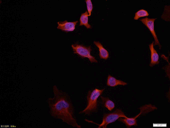
Tissue/Cell: Hela cell, 4% Paraformaldehyde-fixed, Triton X-100 at room temperature for 20 min, Blocking buffer (normal goat serum) at 37°C for 20 min, Antibody incubation with (MAKT1) polyclonal Antibody, Unconjugated (orb11276) 1:100, 90 minutes at 37°C, followed by a conjugated Goat Anti-Mouse IgG-CY3 antibody at 37°C for 90 minutes, DAPI (blue) was used to stain the cell nuclei.
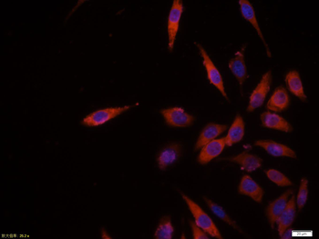
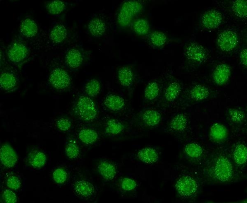
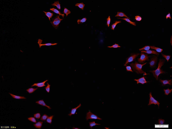
Tissue/Cell: MCF7 cell, 4% Paraformaldehyde-fixed, Triton X-100 at room temperature for 20 min, Blocking buffer (normal goat serum) at 37°C for 20 min, Antibody incubation with (AKT1) polyclonal Antibody, Unconjugated (orb11276) 1:100, 90 minutes at 37°C, followed by a CY3 conjugated Goat Anti-Mouse IgG antibody at 37°C for 90 minutes, DAPI (blue) was used to stain the cell nuclei.
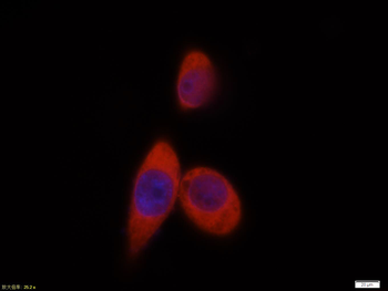
Tissue/Cell: MCF7 cell, 4% Paraformaldehyde-fixed, Triton X-100 at room temperature for 20 min, Blocking buffer (normal goat serum) at 37°C for 20 min, Antibody incubation with (AKT1) polyclonal Antibody, Unconjugated (orb11276) 1:100, 90 minutes at 37°C, followed by a CY3 conjugated Goat Anti-Mouse IgG antibody at 37°C for 90 minutes, DAPI (blue) was used to stain the cell nuclei.
Quick Database Links
Gene Symbol
AKT1
UniProt
UniProt Details
− No UniProt data available
Documents Download
Datasheet
Product Information
Request a Document
Protocol Information
WB
Western Blot (IB, immunoblot)
IHC-P
Immunohistochemistry Paraffin
IHC-Fr
Immunohistochemistry Frozen
IF
Immunofluorescence
ICC
Immunocytochemistry
AKT1 Mouse Polyclonal Antibody (orb11276)

- 0.0
Based on 0 reviews
Participating in our Biorbyt product reviews program enables you to support fellow scientists by sharing your firsthand experience with our products.
Login to Submit a ReviewAvailable Sizes
Select a size below
Free Secondary Antibody (20 ul)0/0
Please add an antibody product to your cart first.