You have no items in your shopping cart.
Description
Research Area
Signal Transduction
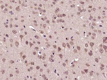
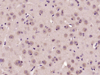
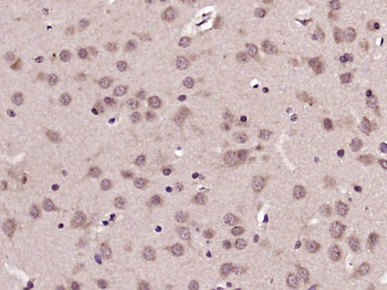
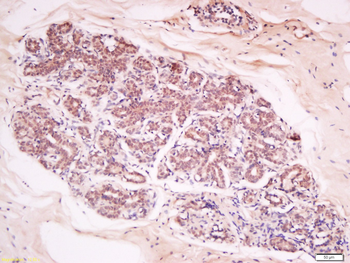
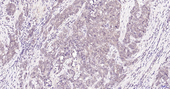
Images & Validation
−Item 1 of 6
| Tested Applications | ELISA, FC, IHC, WB |
|---|---|
| Dilution Range | ELISA: 1:2,000 - 1:10,000, IHC: 20 µg/mL, WB: 1:1000 |
| Reactivity | Human, Mouse |
| Application Notes |
Key Properties
−| Antibody Type | Primary Antibody |
|---|---|
| Host | Mouse |
| Clonality | Monoclonal |
| Isotype | IgG2a |
| Clone No. | 14E5.A2.B2.H9 |
| Immunogen | Anti-AKT1 Antibody was produced by repeated immunizations with a synthetic peptide corresponding to internal residues of human AKT1 protein. |
| Target | AKT1 |
| Purity | Anti-AKT1 antibody is directed against human AKT1. The antibody detects both unphosphorylated and phosphorylated forms of the protein. Anti-AKT1 antibody was purified from ascites by Protein A chromatography. Cross reactivity with AKT1 from other species has not been determined, however, the sequence of the immunogen shows 85% identity to mouse and 92% identity with rat, therefore, cross reactivity is expected. |
| Conjugation | Unconjugated |
Storage & Handling
−| Storage | Store vial at -20° C or below prior to opening. This vial contains a relatively low volume of reagent (25 µL). To minimize loss of volume dilute 1:10 by adding 225 µL of the buffer stated above directly to the vial. Recap, mix thoroughly and briefly centrifuge to collect the volume at the bottom of the vial. Use this intermediate dilution when calculating final dilutions as recommended below. Store the vial at -20°C or below after dilution. Avoid cycles of freezing and thawing. |
|---|---|
| Form/Appearance | Liquid (sterile filtered) |
| Buffer/Preservatives | Preservative: 0.01% (w/v) Sodium Azide. Stabilizer: None; Buffer: 0.02 M Potassium Phosphate, 0.15 M Sodium Chloride, pH 7.2 |
| Concentration | 1.0 mg/ml |
| Expiration Date | 12 months from date of receipt. |
| Dry Ice Shipping | Please note: This product requires shipment on dry ice. A dry ice surcharge will apply. |
| Disclaimer | For research use only |
Alternative Names
−mouse anti-AKT1 antibody, AKT-1, PKB antibody, PKB gamma antibody, PKBGAMMA antibody, PRKBG antibody, Protein kinase Akt 1 antibody, Protein kinase B gamma antibody, RAC-gamma serine/threonine-protein kinase, RAC-PK-gamma
Similar Products
−- Item 1 of 11
Phospho-AKT (Ser473) Rabbit Polyclonal Antibody [orb11293]
WB
Bovine, Canine, Gallus, Mouse, Porcine, Rabbit, Rat, Sheep, Zebrafish
Human
Rabbit
Polyclonal
Unconjugated
50 μl, 100 μl, 200 μl - Item 1 of 9
AKT1 Antibody [orb344403]
ELISA, IF, IHC, Multiplex Assay, WB
Human, Monkey, Mouse, Rat
Mouse
Monoclonal
Unconjugated
100 μg - Item 1 of 11
Phospho-Akt (Thr450) Recombinant Rabbit Monoclonal Antibody [orb559195]
IF, IHC-Fr, IHC-P
Mouse, Rat
Human, Mouse, Rat
Rabbit
Recombinant
Unconjugated
50 μl, 100 μl, 25 μl - Item 1 of 8
AKT1+2+3 Rabbit Polyclonal Antibody [orb155629]
FC, IF, IHC-Fr, IHC-P, WB
Bovine, Canine, Gallus, Porcine, Rabbit, Sheep
Human, Mouse, Rat
Rabbit
Polyclonal
Unconjugated
50 μl, 100 μl, 200 μl - Item 1 of 8
Phospho-Akt1 (Ser473) Recombinant Rabbit Monoclonal Antibody [orb526646]
WB
Mouse, Rat, Zebrafish
Human
Rabbit
Recombinant
Unconjugated
50 μl, 100 μl, 25 μl


Quality Guarantee
Explore bioreagents carefree to elevate your research. All our products are rigorously tested for performance. If a product does not perform as described on its datasheet, our scientific support team will provide expert troubleshooting, a prompt replacement, or a refund. For full details, please see our Terms & Conditions and Buying Guide. Contact us at [email protected].
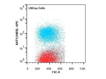
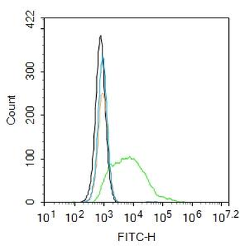
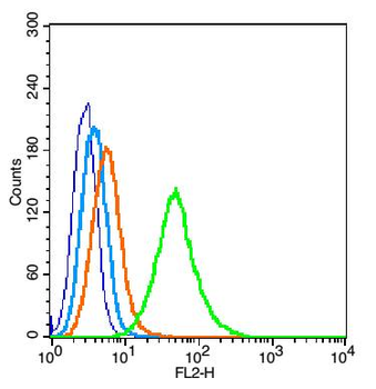
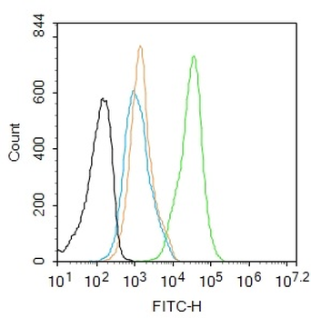
Flow Cytometry of Mouse anti-AKT1 antibody. Cells: LNCap Cells. Stimulation: none. Primary antibody: Allophycocyanin AKT1 antibody at 1.0 µg/ml for 20 min at 4°C.
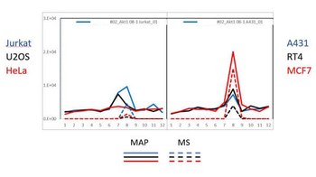
PAGE-MAP (microsphere affinity proteomics) of Mouse Anti-AKT1 Antibody. (orb344523). Antibody array western blot binding of gelfree size separated fractions of multiple lysates (solid lines) and shotgun mass spectroscopy identification (dashed lines) of the target band run in parallel correlate confirming the specificity of this antibody against AKT1.
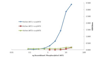
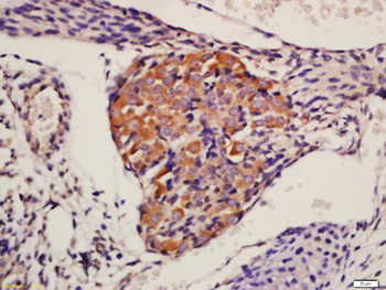
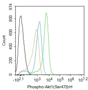
Plate was coated with monoclonal anti AKT1 antibody (capture antibody) followed by incubation with recombinant AKT1 (p/n orb346473), AKT2 (p/n orb346474), AKT3 (p/n orb346475) proteins. Binding was detected with biotinylated monoclonal anti-AKT pS473. The signal shows specificity of the monoclonal anti-AKT1 antibody to recombinant isoform AKT1 protein and not the isoform 2 and 3.
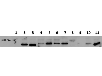
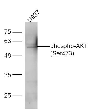
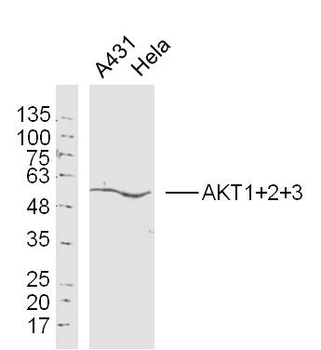
Western Blot of Mouse Anti-AKT1 antibody. Lane 1: AKT-1 Null. Lane 2: WT. Lane 3: MEF #1. Lane 4: A549. Lane 5: Calu-1. Lane 6: PC-3 (p/n orb692718). Lane 7: HepG2 (p/n orb348735). Lane 8: Jurkat (p/n orb348674). Lane 9: SKOV3. Lane 10: HEK293T. Lane 11: C2C12 (p/n orb348725). Load: 20 ug per lane. Primary antibody: AKT1 antibody at 1:1000 for overnight at 4°C. Secondary antibody: Peroxidase mouse secondary antibody at 1:40000 for 30 min at RT. Block: orb348637 for 30 min at RT. Predicted/Observed size: 56 kDa for AKT1. Other band(s): none.

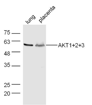
Western Blot of Mouse Anti-AKT1 antibody. Lane 1: GST Tagged recombinant AKT1. Lane 2: GST Tagged recombinant AKT2. Lane 3: GST Tagged recombinant AKT3. Load: 25 ng per lane. Primary antibody: AKT1 antibody at 1:1000 for overnight at 4°C. Secondary antibody: Peroxidase mouse secondary antibody at 1:40000 for 30 min at RT. Block: orb348637 for 30 min at RT. Predicted/Observed size: 78 kDa for AKT1. Other band(s): none.
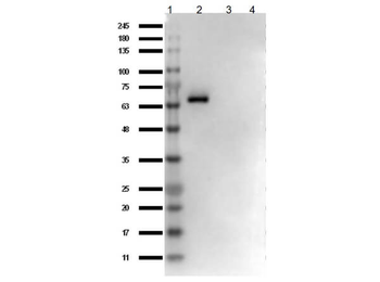
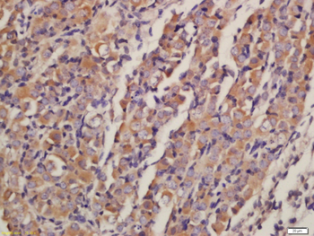
Western Blot of Mouse Anti-AKT1 Antibody. Lane 1: Opal Prestained Molecular Weight Protein. Lane 2: AKT1 protein (p/n orb346473). Lane 3: AKT2 protein (p/n orb346474). Lane 4: AKT3 protein (p/n orb346475). Load: 50 ng. Blocking: BlockOut Buffer (p/n orb348644) for 30 min at RT. Primary Antibody: Anti-AKT1 at 1 ug/ml o/n at 4°C. Secondary Antibody: Rabbit Anti-Mouse IgG HRP (p/n orb347506) at 1:40000 in orb348644 for 30 min at RT.
Quick Database Links
Gene Symbol
AKT1
UniProt
UniProt Details
− No UniProt data available
Documents Download
Datasheet
Product Information
Request a Document
Protocol Information
WB
Western Blot (IB, immunoblot)
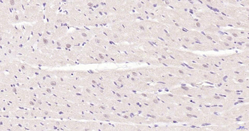
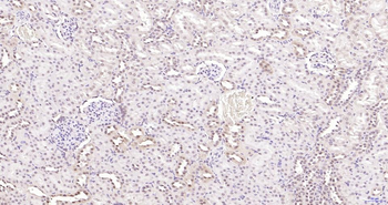
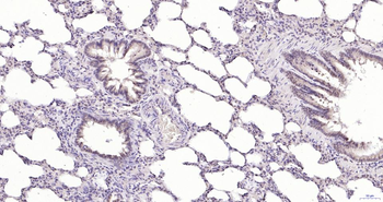
IHC
Immunohistochemistry
FC
Flow Cytometry
ELISA
Enzyme-linked Immunosorbent Assay (EIA)
AKT1 Antibody (orb344525)

- 0.0
Based on 0 reviews
Participating in our Biorbyt product reviews program enables you to support fellow scientists by sharing your firsthand experience with our products.
Login to Submit a ReviewAvailable Sizes
Select a size below
Free Secondary Antibody (20 ul)0/0
Please add an antibody product to your cart first.