You have no items in your shopping cart.
Description
Research Area
Signal Transduction
Images & Validation
−Item 1 of 4
| Tested Applications | FC, WB |
|---|---|
| Dilution Range | WB - 1:1000, FC - 1:10-50 |
| Reactivity | Human |
| Predicted Reactivity | Mouse, Rat |
Key Properties
−| Host | Rabbit |
|---|---|
| Clonality | Polyclonal |
| Isotype | Rabbit IgG |
| Immunogen | This AKT1 antibody is generated from rabbits immunized with a KLH conjugated synthetic peptide between 438-468 amino acids from the C-terminal region of human AKT1. Antigen Region: 438-468 aa. |
| Target | AKT1 |
| Molecular Weight | 55686 Da |
| Conjugation | Unconjugated |
Storage & Handling
−| Storage | Maintain refrigerated at 2-8°C for up to 2 weeks. For long term storage store at -20°C in small aliquots to prevent freeze-thaw cycles |
|---|---|
| Form/Appearance | Purified polyclonal antibody supplied in PBS with 0.09% (W/V) sodium azide. This antibody is prepared by Saturated Ammonium Sulfate (SAS) precipitation followed by dialysis against PBS. |
| Expiration Date | 12 months from date of receipt. |
| Disclaimer | For research use only |
Alternative Names
−RAC-alpha serine/threonine-protein kinase, Protein kinase B, PKB, Protein kinase B alpha, PKB alpha, Proto-oncogene c-Akt, RAC-PK-alpha, AKT1, PKB, RAC
Similar Products
−- Item 1 of 5
AKT1 Antibody (C-term T450) [orb1438858]
FC, IF, IHC-P, WB
Rat
Human, Mouse
Rabbit
Polyclonal
Unconjugated
50 μl, 100 μl - Item 1 of 2
- Item 1 of 2
AKT Rabbit Polyclonal Antibody [orb224167]
IHC, WB
Bovine, Human, Mouse, Rat, Sheep, Zebrafish
Rabbit
Polyclonal
Unconjugated
30 μl, 100 μl, 200 μl, 50 μl - Item 1 of 2
PRAS40 Rabbit Polyclonal Antibody [orb304683]
IHC, WB
Bovine, Human, Mouse, Rat
Rabbit
Polyclonal
Unconjugated
30 μl, 100 μl, 200 μl, 50 μl
Quality Guarantee
Explore bioreagents carefree to elevate your research. All our products are rigorously tested for performance. If a product does not perform as described on its datasheet, our scientific support team will provide expert troubleshooting, a prompt replacement, or a refund. For full details, please see our Terms & Conditions and Buying Guide. Contact us at [email protected].
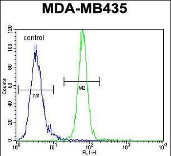
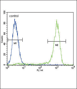
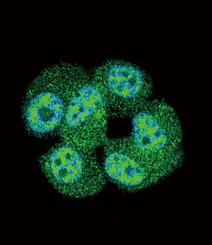
AKT1 Antibody (C-term) flow cytometric analysis of MDA-MB435 cells (right histogram) compared to a negative control cell (left histogram). FITC-conjugated goat-anti-rabbit secondary antibodies were used for the analysis.
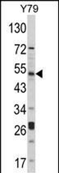
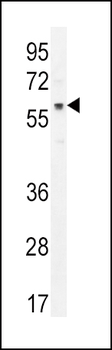
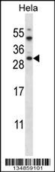
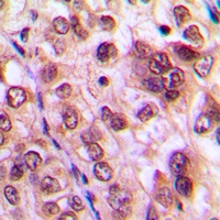
Western blot analysis of hAKT1-D453 in Y79 cell line lysates (35 ug/lane).AKT1 (arrow) was detected using the purified Pab.
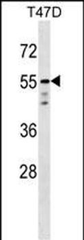
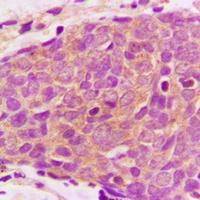
AKT1 Antibody (C-term) western blot analysis in T47D cell line lysates (35 ug/lane). This demonstrates the AKT1 antibody detected the AKT1 protein (arrow).
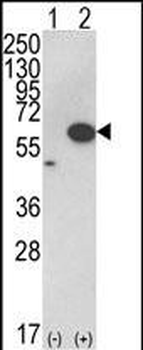
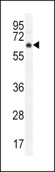
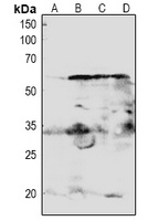
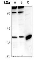
Western blot analysis of AKT1 (arrow) using rabbit polyclonal hAKT1-D453. 293 cell lysates (2 ug/lane) either nontransfected (Lane 1) or transiently transfected with the AKT1 gene (Lane 2).
Quick Database Links
UniProt Details
− No UniProt data available
NCBI Reference Sequences
−Associated Accession Numbers
Curated reference sequences for the gene transcript and protein product| Protein | NP_001014432.1, NP_001014431.1, NP_005154.2 |
|---|
Documents Download
Datasheet
Product Information
Request a Document
Protocol Information
WB
Western Blot (IB, immunoblot)
FC
Flow Cytometry
AKT1 Antibody (C-term) (orb1929633)

- 0.0
Based on 0 reviews
Participating in our Biorbyt product reviews program enables you to support fellow scientists by sharing your firsthand experience with our products.
Login to Submit a ReviewAvailable Sizes
Select a size below
Choose Conjugation or Carrier Free Version
Free Secondary Antibody (20 ul)0/0
Please add an antibody product to your cart first.