You have no items in your shopping cart.
Description
Research Area
Signal Transduction
Images & Validation
−Item 1 of 8
| Tested Applications | DOT, ELISA, IHC, WB |
|---|---|
| Dilution Range | ELISA: 1:15,000, IHC: 1:200, WB: 1:1000 |
| Reactivity | Human, Mouse, Rat |
| Application Notes |
Key Properties
−| Antibody Type | Primary Antibody |
|---|---|
| Host | Rabbit |
| Clonality | Polyclonal |
| Isotype | IgG |
| Immunogen | Anti-AKT pT308 polyclonal antibody was produced by repeated immunizations with a phosphorylated synthetic peptide corresponding to residues surrounding threonine 308 of human AKT1 protein. |
| Target | AKT1 |
| Purity | AKT phospho T308 Antibody was prepared from monospecific antiserum by immunoaffinity chromatography using phospho peptide coupled to agarose beads followed by solid phase adsorption(s) against non-phospho peptide and non-specific peptide to remove any unwanted reactivities. Assay by immunoelectrophoresis resulted in a single precipitin arc against anti-Rabbit Serum. This antibody is specific for phosphorylated human AKT. Minimal reactivity occurs against non-phosphorylated AKT. Reactivity against AKT from other species may occur but has not yet been tested. |
| Conjugation | Unconjugated |
Storage & Handling
−| Storage | Store AKT phospho T308 Antibody at -20° C prior to opening. Aliquot contents and freeze at -20° C or below for extended storage. Avoid cycles of freezing and thawing. Centrifuge product if not completely clear after standing at room temperature. This product is stable for several weeks at 4° C as an undiluted liquid. Dilute only prior to immediate use. |
|---|---|
| Form/Appearance | Liquid (sterile filtered) |
| Buffer/Preservatives | Preservative: 0.01% (w/v) Sodium Azide. Stabilizer: None; Buffer: 0.02 M Potassium Phosphate, 0.15 M Sodium Chloride, pH 7.2 |
| Concentration | 1.0 mg/mL |
| Expiration Date | 12 months from date of receipt. |
| Dry Ice Shipping | Please note: This product requires shipment on dry ice. A dry ice surcharge will apply. |
| Disclaimer | For research use only |
Alternative Names
−rabbit anti-AKT pT308 Antibody, AKT1 phospho T308, RAC-PK-alpha, Protein kinase B, PKB, C-AKT, RAC-alpha serine/threonine-protein kinase, Proto-oncogene c-Akt, AKT1, AKT 1, AKT-1
Similar Products
−- Item 1 of 11
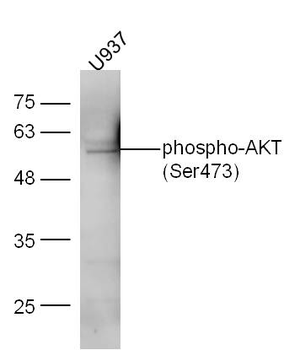
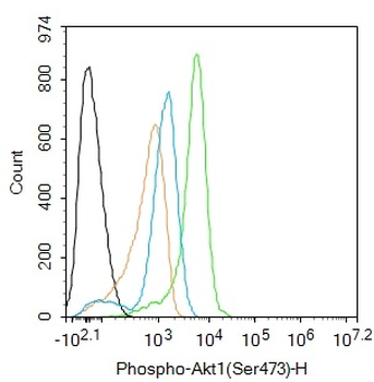
Phospho-AKT (Ser473) Rabbit Polyclonal Antibody [orb11293]
WB
Bovine, Canine, Gallus, Mouse, Porcine, Rabbit, Rat, Sheep, Zebrafish
Human
Rabbit
Polyclonal
Unconjugated
50 μl, 100 μl, 200 μl - Item 1 of 9
AKT1 Antibody [orb344403]
ELISA, IF, IHC, Multiplex Assay, WB
Human, Monkey, Mouse, Rat
Mouse
Monoclonal
Unconjugated
100 μg - Item 1 of 11
Phospho-Akt (Thr450) Recombinant Rabbit Monoclonal Antibody [orb559195]
IF, IHC-Fr, IHC-P
Mouse, Rat
Human, Mouse, Rat
Rabbit
Recombinant
Unconjugated
50 μl, 100 μl, 25 μl - Item 1 of 8
AKT1+2+3 Rabbit Polyclonal Antibody [orb155629]
FC, IF, IHC-Fr, IHC-P, WB
Bovine, Canine, Gallus, Porcine, Rabbit, Sheep
Human, Mouse, Rat
Rabbit
Polyclonal
Unconjugated
50 μl, 100 μl, 200 μl - Item 1 of 8
Phospho-Akt1 (Ser473) Recombinant Rabbit Monoclonal Antibody [orb526646]
WB
Mouse, Rat, Zebrafish
Human
Rabbit
Recombinant
Unconjugated
50 μl, 100 μl, 25 μl


Quality Guarantee
Explore bioreagents carefree to elevate your research. All our products are rigorously tested for performance. If a product does not perform as described on its datasheet, our scientific support team will provide expert troubleshooting, a prompt replacement, or a refund. For full details, please see our Terms & Conditions and Buying Guide. Contact us at [email protected].
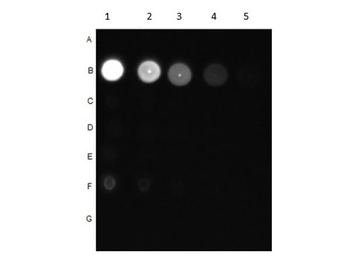
Dot Blot of Rabbit Anti-AKT pT308 Antibody. Dilutions in Columns: (1) 100 ng, (2) 33.33 ng, (3) 11.11 ng, (4) 3.7 ng, (5) 1.23 ng. Tested BSA Peptide Reactivity in Rows: (A) AKT1-BSA, (B) AKT1 pT308-BSA, (C) AKT1 S473-BSA, (D) AKT1 pS473-BSA, (E) CDC27 T244-BSA, (F) CDC27 pT244-BSA, (G) BSA control. Primary Antibody: Anti-AKT pT308 at 1 µg/ml overnight at 2-8°C. Secondary Antibody: Goat anti-Rabbit IgG HRP (p/n orb347654) at 1:70000 at RT for 30 mins. Block: BlockOut Buffer (p/n orb348644).
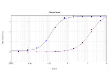
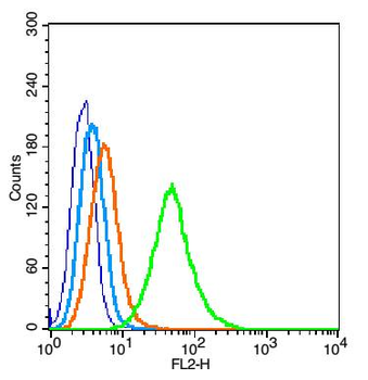
ELISA Results of Rabbit Anti-AKT pT308 Antibody tested against BSA-conjugated non-phospho [purple] and phospho [blue] forms of immunizing peptide. Each well was coated in duplicate with either 0.1 µg of conjugate. The working dilution is 1:81300. The starting dilution of antibody was 5 µg/ml and the X-axis represents the Log10 of a 3-fold dilution. This titration is a 4-parameter curve fit where the IC50 is defined as the titer of the antibody. Assay performed using HRP conjugate stabilizer, Goat Anti-Rabbit HRP conjugated (p/n orb347654) and TMB substrate (p/n orb348651).
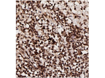
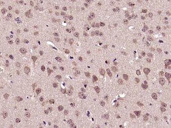
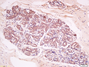
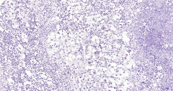
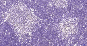
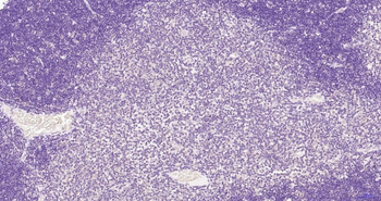
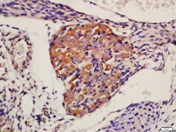
Immunohistochemistry of Rabbit Anti-AKT pT308 Antibody. Tissue: human breast tissue (lymph nodes). Antigen retrieval: HIER using citrate buffer for 20 minutes. Fixative: None. Primary Antibody: Anti-AKT phosphoT308 at 1:200 for 30 minutes at RT. Secondary Antibody: Anti-Rabbit Poly-HRP-IgG Ready to Use for 8 minutes at RT. Counterstain: Hematoxylin. Substrate: DAB. Analysis: Strong staining in nucleus. May be suitable with more dilutions.
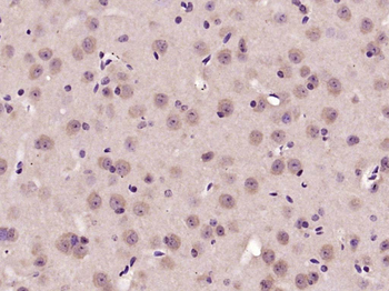
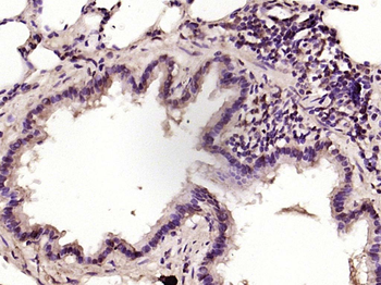
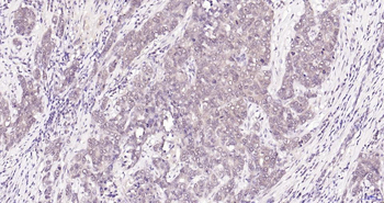
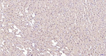
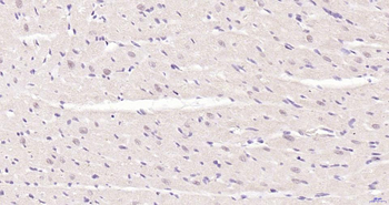
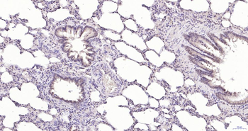
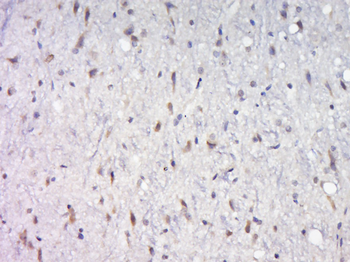
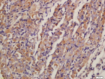
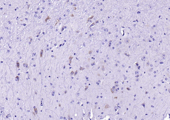
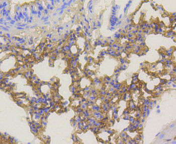
Immunohistochemistry of Rabbit Anti-AKT pT308 Antibody. Tissue: human lung tissue. Antigen retrieval: HIER using citrate buffer for 20 minutes. Fixative: None. Primary Antibody: Anti-AKT phosphoT308 at 1:200 for 30 minutes at RT. Secondary Antibody: Anti-Rabbit Poly-HRP-IgG Ready to Use for 8 minutes at RT. Counterstain: Hematoxylin. Substrate: DAB. Analysis: Strong staining in nucleus. May be suitable with more dilutions.
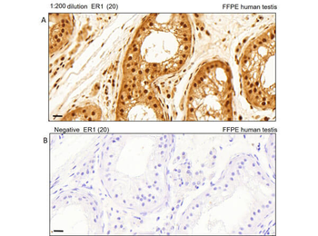
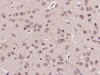
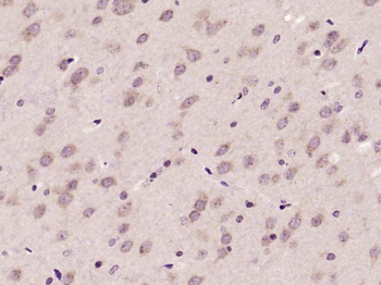
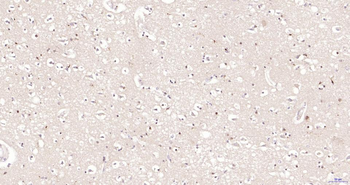
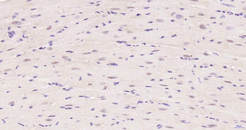
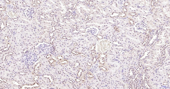
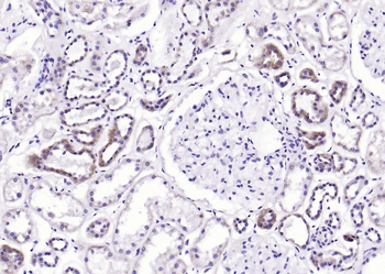
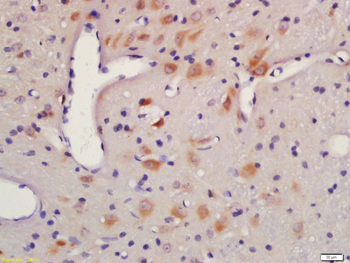
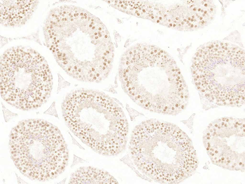
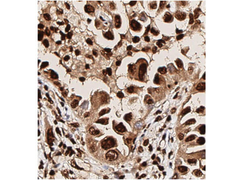
Immunohistochemistry of Rabbit Anti-AKT pT308 Antibody. Tissue: human testis tissue. Antigen retrieval: Heat induced antigen retrieval was performed using Leica Bond Epitope Retrieval Buffer 1 (Citrate solution, pH6.0) for 20 minutes. Fixative: None. Primary Antibody: (A). Anti-AKTpT308 at 1:200 for 30 minutes at RT. (B). Negative control. Secondary Antibody: Anti-Rabbit Poly-HRP-IgG Ready to Use for 8 minutes at RT. Counterstain: Hematoxylin. Substrate: DAB. Analysis: Cells in seminiferous ducts and Leydig cells show moderate cytoplasmic staining.
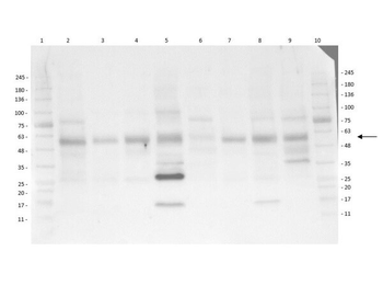
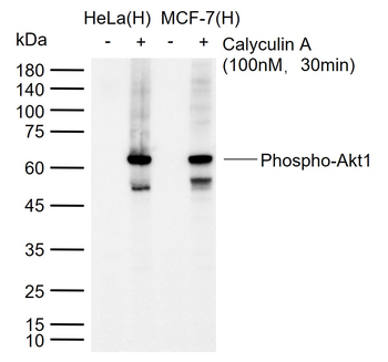
Multi-Lysate Western Blot of Rabbit Anti-AKT pT308 Antibody. Lane 1: Opal Pre-stained Molecular Weight Marker. Lane 2: Human Spleen Lysate. Lane 3: Hu Small Intestine Lysate. Lane 4: Hu Placenta Lysate. Lane 5: Hu Skeletal Muscle Lysate. Lane 6: Hu Brain Cerebellum Lysate. Lane 7: Hu Lung Lysate. Lane 8: Hu Tonsil Lysate. Lane 9: Hu Thymus Lysate. Lane 10: Opal Pre-stained Molecular Weight Marker. Primary Antibody: Anti-AKT pT308 at 1:1000 overnight at 2-8°C. Secondary Antibody: Goat Anti-Rabbit IgG HRP (p/n orb347654) at 1:40000 at RT for 60 mins. Block: BlockOut Buffer (p/n orb348644). Predicted MW: ~55kDa. Observed MW: ~28, ~58kDa. Notes: Ubiquitous.
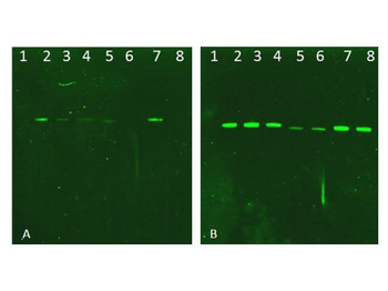
Western Blot of Rabbit AKT Antibodies. Lane 1: NIR MW protein ladder. Lane 2: AKT1, recombinant: orb346473. Lane 3: AKT1, phosphatase-treated: orb346472. Lane 4: AKT1, mutant T308A/S473A: orb346474. Lane 5: AKT2, recombinant: orb346475. Lane 6: AKT2, phosphatase-treated: orb346470. Lane 7: AKT3, recombinant: orb346476. Lane 8: AKT3, phosphatase-treated: orb346471. Load: 50 ng per lane. Blot A: orb345379 Anti-Akt pT308 used at 1:2270, Blot B: orb750474 Anti-Akt used 1:1000.
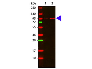
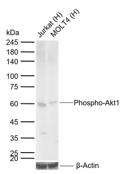
Western Blot of Rabbit anti-Akt phospho T308 antibody. Lane 1: GST tagged AKT1 un-active recombinant protein. Lane 2: GST tagged AKT1 active recombinant protein. Load: 50 ng per lane. Primary antibody: Akt phospho T308 antibody at 1:1000 for overnight at 4°C. Secondary antibody: DyLight™ 649 rabbit secondary antibody at 1:20000 for 30 min at RT. Block: orb348637 for 30 min at RT. Other band(s): none.
Documents Download
Datasheet
Product Information
Request a Document
Protocol Information
WB
Western Blot (IB, immunoblot)
IHC
Immunohistochemistry
ELISA
Enzyme-linked Immunosorbent Assay (EIA)
DOT
Dot Blot
AKT1 Antibody (orb345379)

- 0.0
Based on 0 reviews
Participating in our Biorbyt product reviews program enables you to support fellow scientists by sharing your firsthand experience with our products.
Login to Submit a ReviewAvailable Sizes
Select a size below
Choose Conjugation or Carrier Free Version
Free Secondary Antibody (20 ul)0/0
Please add an antibody product to your cart first.