You have no items in your shopping cart.
Description
Research Area
Signal Transduction
Images & Validation
−Item 1 of 7
| Tested Applications | ELISA, IF, IHC, WB |
|---|---|
| Dilution Range | ELISA: 1:20,000, IHC: 20 µg/ml, IF: 1:500 - 1:3,000, WB: 1:500 - 1:3,000 |
| Reactivity | Human, Mouse |
| Application Notes |
Key Properties
−| Antibody Type | Primary Antibody |
|---|---|
| Host | Mouse |
| Clonality | Monoclonal |
| Isotype | IgG1 |
| Clone No. | 17F6.B11 |
| Immunogen | This monoclonal antibody was produced by repeated immunizations with a synthetic peptide corresponding to residues surrounding S473 of human AKT1 protein. |
| Target | AKT1 |
| Purity | This product was purified from concentrated tissue culture supernate by Protein A chromatography. This antibody is specific for human and mouse AKT protein phosphorylated at S473. A BLAST analysis was used to suggest cross-reactivity with AKT pS473 from human, mouse, rat and chimpanzee sources based on 100% homology with the immunizing sequence. Cross-reactivity with AKT from other sources has not been determined. Cross-reactivity with AKT2 and AKT3 has not been determined. |
| Conjugation | Unconjugated |
Storage & Handling
−| Storage | Store vial at -20° C prior to opening. Aliquot contents and freeze at -20° C or below for extended storage. Avoid cycles of freezing and thawing. Centrifuge product if not completely clear after standing at room temperature. This product is stable for several weeks at 4° C as an undiluted liquid. Dilute only prior to immediate use. |
|---|---|
| Form/Appearance | Liquid (sterile filtered) |
| Buffer/Preservatives | Preservative: 0.01% (w/v) Sodium Azide. Stabilizer: None; Buffer: 0.02 M Potassium Phosphate, 0.15 M Sodium Chloride, pH 7.2 |
| Concentration | 1.0 mg/mL |
| Expiration Date | 12 months from date of receipt. |
| Dry Ice Shipping | Please note: This product requires shipment on dry ice. A dry ice surcharge will apply. |
| Disclaimer | For research use only |
Alternative Names
−mouse anti-AKT pS473 Antibody, RAC-PK-alpha, Protein kinase B, PKB, C-AKT, RAC-alpha serine/threonine-protein kinase, Proto-oncogene c-Akt, AKT1, AKT 1, AKT-1
Similar Products
−- Item 1 of 11
Phospho-AKT (Ser473) Rabbit Polyclonal Antibody [orb11293]
WB
Bovine, Canine, Gallus, Mouse, Porcine, Rabbit, Rat, Sheep, Zebrafish
Human
Rabbit
Polyclonal
Unconjugated
50 μl, 100 μl, 200 μl - Item 1 of 9
AKT1 Antibody [orb344403]
ELISA, IF, IHC, Multiplex Assay, WB
Human, Monkey, Mouse, Rat
Mouse
Monoclonal
Unconjugated
100 μg - Item 1 of 11
Phospho-Akt (Thr450) Recombinant Rabbit Monoclonal Antibody [orb559195]
IF, IHC-Fr, IHC-P
Mouse, Rat
Human, Mouse, Rat
Rabbit
Recombinant
Unconjugated
50 μl, 100 μl, 25 μl - Item 1 of 8
AKT1+2+3 Rabbit Polyclonal Antibody [orb155629]
FC, IF, IHC-Fr, IHC-P, WB
Bovine, Canine, Gallus, Porcine, Rabbit, Sheep
Human, Mouse, Rat
Rabbit
Polyclonal
Unconjugated
50 μl, 100 μl, 200 μl - Item 1 of 8
Phospho-Akt1 (Ser473) Recombinant Rabbit Monoclonal Antibody [orb526646]
WB
Mouse, Rat, Zebrafish
Human
Rabbit
Recombinant
Unconjugated
50 μl, 100 μl, 25 μl


Quality Guarantee
Explore bioreagents carefree to elevate your research. All our products are rigorously tested for performance. If a product does not perform as described on its datasheet, our scientific support team will provide expert troubleshooting, a prompt replacement, or a refund. For full details, please see our Terms & Conditions and Buying Guide. Contact us at [email protected].
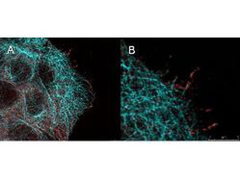
High resolution STED immunofluorescence nanoscopy of Mouse anti-AKT pS473 antibody. Tissue: A431 cells. The merge images (A) and at high magnification (B) show phosphorylated AKT colocalized with the distal microtubules. Fixation: 4% paraformaldehyde for 5 min and after washes blocked with 10% NGS/0.2% Triton X-100 for 30 min. Antigen retrieval: serum deprivation for 12 h. Primary antibody: AKT pS473 antibody at 10 µg/ml and α-tubulin (cyan) (p/n orb345510) at 1.4 µg/ml for 1 h at RT. Secondary antibody: Atto 647N anti-Mouse IgG (ATTO TEC GmbH), and DyLight™488 anti-Rabbit IgG were used at 1.0 µg/ml for 1h at RT for indirect detection. Localization: AKT pS473 is in the cytoplasm and also organized at the periphery of the cell. Staining: AKT pS473 as red signal with bis-benzimide (blue) nuclear counterstain.
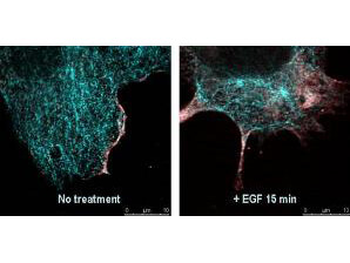
Immunofluorescence confocal microscopy of Mouse Anti-AKT pS473 antibody. Tissue: EGF treated A431 cells. Fixation: 0.5% PFA. Antigen retrieval: EGF 15 min. Primary antibody: AKT pS473 antibody at 10 µg/ml for 1 h at RT. Secondary antibody: DyLight 488™ Goat anti-Rabbit IgG, MAb anti-AKT pS473, atto-647N anti-Mouse IgG (Active Motif). at 1:10000 for 45 min at RT. Localization: AKT pS473 is nuclear and occasionally cytoplasmic. Staining: AKT pS473 as red signal with tubulin (cyan).
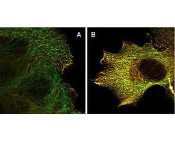
Immunofluorescence Microscopy of Mouse Anti-AKTpS473 antibody using STED nanoscopy to evaluate AKT activation and migration. Tissue: A431 cells. Antigen retrieval: Panel A: serum starved, unstimulated cells. Panel B: serum starved, EGF stimulated for 15 mins. A massive increase in AKT-pS473 activation, as measured by intensity signal, peaked at 15 minutes and was associated with depolymerized tubulin. Staining: Panel A shows STED data (AKT-pS473, red channel) collected simultaneously with confocal signal (a-tubulin, green channel). Upon stimulation of cells with EGF, a rapid activation of AKT is observed (Panel B) along with a coincident change in the tubulin organization (yellow signal), as well as an extensive cell shape-change (cell membrane folding) and accumulation of AKTpS473 at the cell periphery.
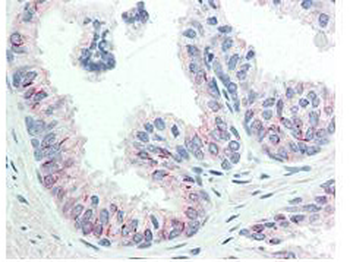
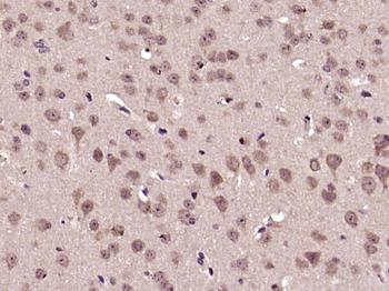
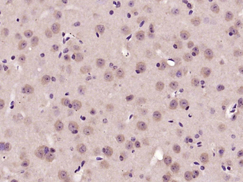
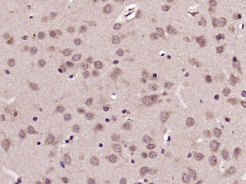
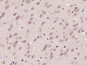
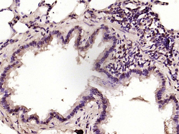
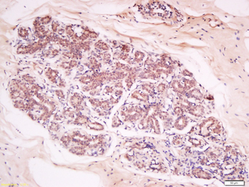
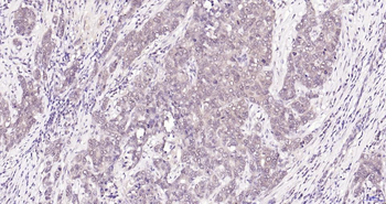
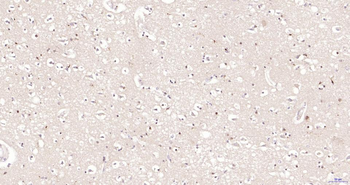
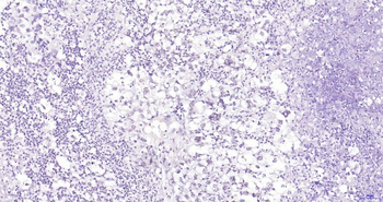
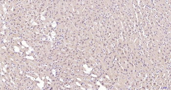
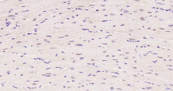
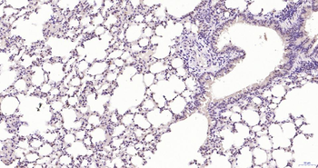
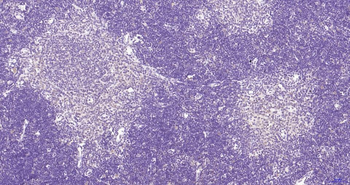
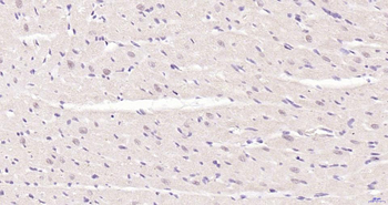
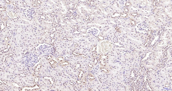
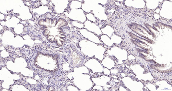
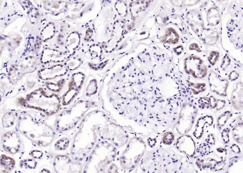
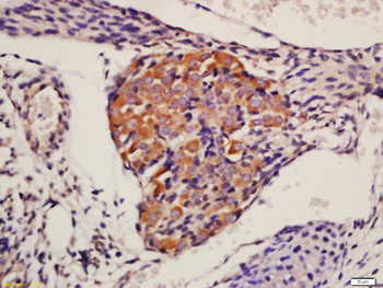
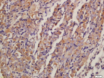
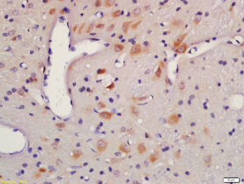
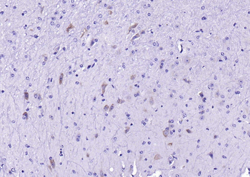
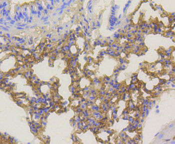
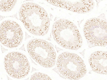
Immunohistochemistry of Mouse anti-AKT pS473 antibody. Tissue: human prostate tissue. Fixation: formalin fixed paraffin embedded. Antigen retrieval: not required. Primary antibody: AKT pS473 antibody at 20 µg/ml for 1 h at RT. Secondary antibody: Dako's Techmate streptavidin-biotin reagents at 1:10000 for 45 min at RT. Localization: AKT pS473 is nuclear and occasionally cytoplasmic. Staining: AKT pS473 as precipitated red signal with hematoxylin purple nuclear counterstain.
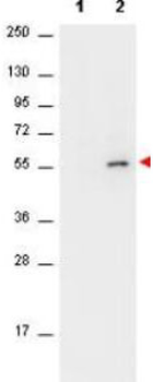
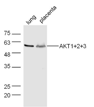
Western Blot of Mouse anti-AKT antibody. Lane 1: unstimulated NIH/3T3 cell lysates (p/n orb348714). Lane 2: PDGF stimulated NIH/3T3 cell lysates (p/n orb348723). Load: 10 µg per lane. Primary antibody: AKT antibody at 1:400 for overnight at 4°C. Secondary antibody: HRP conjugated Gt-a-Mouse IgG (p/n orb347385) was used at a 1:40000 dilution for 1 h at 4°C with FemtoMax™ enhanced chemiluminescent reagent. Block: 5% BLOTTO (p/n orb348624) in TBS for 2h at RT. Observed size: ~56 kDa for AKT. Other band(s): none.
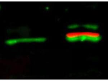
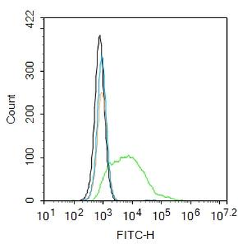
Western Blot of Mouse Anti-Akt pS473 antibody. Lane 1: unstimulated NIH/3T3 lysates (p/n orb348714) contain inactive unphosphorylated Akt1, green band. Lane 2: PDGF stimulated NIH/3T3 lysate (p/n orb348723) contains both inactive (green band) and activated phosphorylated Akt1 (red band). Load: 10 µg per lane. Primary antibody: rabbit anti-Akt (pan) (p/n orb750474) and mouse anti-Akt pS473 (p/n orb344456) specific antibodies at 1:400 for overnight at 4°C. Secondary antibody: anti-rabbit IgG DyLight™ 549 (green) and anti-mouse IgG DyLight™ 649 conjugated (red) secondary antibodies at 1:10000 for 45 min at RT. Block: 5% BLOTTO overnight at 4°C.
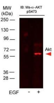
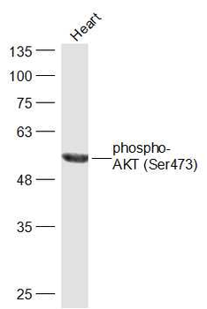
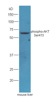
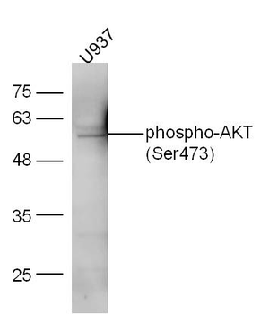
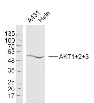
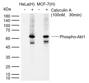
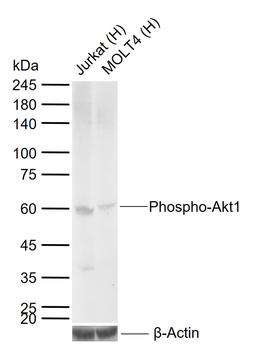
Western Blot of Mouse Anti-AKTpS473 antibody. Lane 1: A431 cell lysate (p/n orb348665). Lane 2: A431 cells stimulated for 15 min with EGF (p/n orb348666). Load: 35 µg per lane. Primary antibody: AKTpS473 antibody at 1:400 for overnight at 4°C. Secondary antibody: DyLight™649 Conjugated Anti-AKT pS473 Monoclonal Antibody at 1:10000 for 45 min at RT. Block: Blocking Buffer for Fluorescent Western Blotting (p/n orb348637) overnight at 4°C. Predicted/Observed size: 56kDa. Other band(s): none.
Documents Download
Datasheet
Product Information
Request a Document
Protocol Information
WB
Western Blot (IB, immunoblot)
IHC
Immunohistochemistry
IF
Immunofluorescence
ELISA
Enzyme-linked Immunosorbent Assay (EIA)
AKT1 Antibody (orb344456)

- 0.0
Based on 0 reviews
Participating in our Biorbyt product reviews program enables you to support fellow scientists by sharing your firsthand experience with our products.
Login to Submit a ReviewAvailable Sizes
Select a size below
Free Secondary Antibody (20 ul)0/0
Please add an antibody product to your cart first.