You have no items in your shopping cart.
Description
Research Area
Signal Transduction
Images & Validation
−Item 1 of 7
| Tested Applications | ELISA, IF, IHC, WB |
|---|---|
| Dilution Range | ELISA: 1:2,000 - 1:10,000, IHC: 1:500 - 1:2,000, IF: 1:100 - 1:1,000, WB: 1:500 - 1:2,000 |
| Reactivity | Gallus, Human, Mouse, Rat |
| Application Notes |
Key Properties
−| Antibody Type | Primary Antibody |
|---|---|
| Host | Rabbit |
| Clonality | Polyclonal |
| Isotype | Antiserum |
| Immunogen | AKT Antibody was produced from whole rabbit serum prepared by repeated immunizations with a synthetic peptide R-P-H-F-P-Q-F-S-Y-S-A-S-G-T-A corresponding to the C-terminus (460-480) of human AKT proteins conjugated to KLH using maleimide. A residue of cysteine was added to the amino terminal end to facilitate coupling. A BLAST analysis was used to suggest reactivity with this protein from rat, mouse, and chicken based on 100% homology for the immunogen sequence. |
| Target | AKT1 |
| Purity | This product was prepared from monospecific antiserum by a delipidation and defibrination. Pan Anti-AKT Antibody reacts with the AKT from human tissues. Based on sequence we expect this antibody to react as well with rat, mouse, and chicken AKT. |
| Conjugation | Unconjugated |
Storage & Handling
−| Storage | Store vial at -20° C or below prior to opening. This vial contains a relatively low volume of reagent (25 µL). To minimize loss of volume dilute 1:10 by adding 225 µL of the buffer stated above directly to the vial. Recap, mix thoroughly and briefly centrifuge to collect the volume at the bottom of the vial. Use this intermediate dilution when calculating final dilutions as recommended below. Store the vial at -20°C or below after dilution. Avoid cycles of freezing and thawing. |
|---|---|
| Form/Appearance | Liquid (sterile filtered) |
| Buffer/Preservatives | 0.1% (w/v) Sodium Azide |
| Concentration | 75 mg/ml |
| Expiration Date | 12 months from date of receipt. |
| Dry Ice Shipping | Please note: This product requires shipment on dry ice. A dry ice surcharge will apply. |
| Disclaimer | For research use only |
Alternative Names
−rabbit anti-AKT antibody, RAC-PK-alpha, Protein kinase B, PKB, C-AKT, RAC-alpha serine/threonine-protein kinase, Proto-oncogene c-Akt, AKT1, AKT 1, AKT-1
Similar Products
−- Item 1 of 11
Phospho-AKT (Ser473) Rabbit Polyclonal Antibody [orb11293]
WB
Bovine, Canine, Gallus, Mouse, Porcine, Rabbit, Rat, Sheep, Zebrafish
Human
Rabbit
Polyclonal
Unconjugated
50 μl, 100 μl, 200 μl - Item 1 of 9
AKT1 Antibody [orb344403]
ELISA, IF, IHC, Multiplex Assay, WB
Human, Monkey, Mouse, Rat
Mouse
Monoclonal
Unconjugated
100 μg - Item 1 of 11
Phospho-Akt (Thr450) Recombinant Rabbit Monoclonal Antibody [orb559195]
IF, IHC-Fr, IHC-P
Mouse, Rat
Human, Mouse, Rat
Rabbit
Recombinant
Unconjugated
50 μl, 100 μl, 25 μl - Item 1 of 8
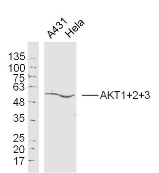
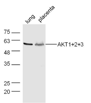
AKT1+2+3 Rabbit Polyclonal Antibody [orb155629]
FC, IF, IHC-Fr, IHC-P, WB
Bovine, Canine, Gallus, Porcine, Rabbit, Sheep
Human, Mouse, Rat
Rabbit
Polyclonal
Unconjugated
50 μl, 100 μl, 200 μl - Item 1 of 8
Phospho-Akt1 (Ser473) Recombinant Rabbit Monoclonal Antibody [orb526646]
WB
Mouse, Rat, Zebrafish
Human
Rabbit
Recombinant
Unconjugated
50 μl, 100 μl, 25 μl


Quality Guarantee
Explore bioreagents carefree to elevate your research. All our products are rigorously tested for performance. If a product does not perform as described on its datasheet, our scientific support team will provide expert troubleshooting, a prompt replacement, or a refund. For full details, please see our Terms & Conditions and Buying Guide. Contact us at [email protected].
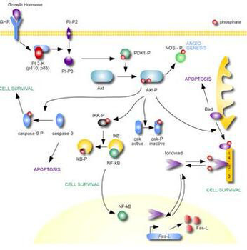
AKT Metabolic Pathway
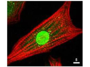
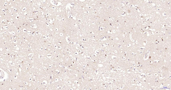
Immunofluorescence Microscopy of Rabbit Anti-AKT Antibody. Tissue: neonatal rat cardiomyocytes. Fixation: 0.5% PFA. Antigen retrieval: not required. Primary antibody: AKT antibody at 1:80 dilution for 1 h at RT. Secondary antibody: Texas-red™ conjugated rabbit secondary antibody at 1:10000 for 45 min at RT. Localization: AKT is nuclear. Staining: Anti-AKT staining appears green. Actin filaments are labeled red using a Texas-red™ conjugated phalloidin.
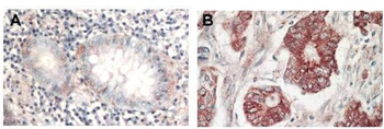
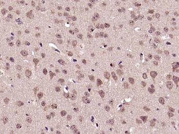
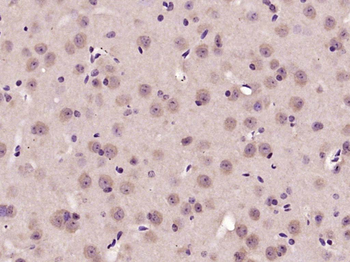
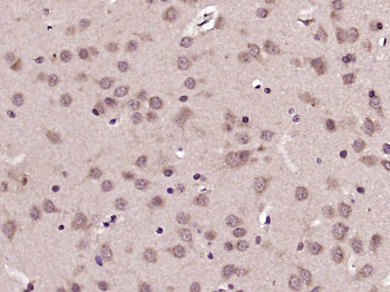
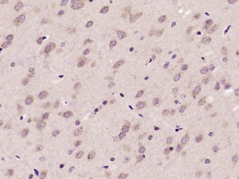
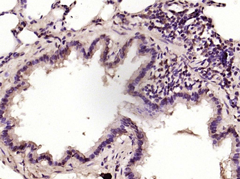
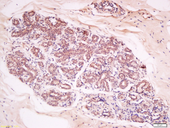
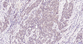
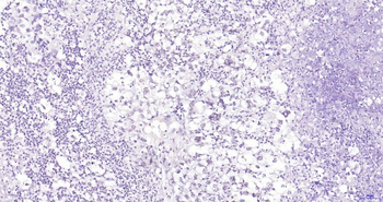
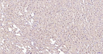
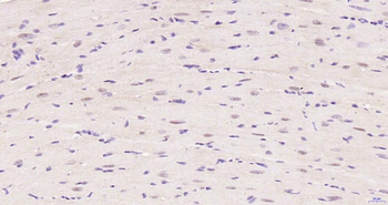
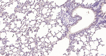
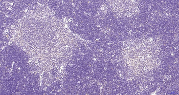
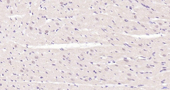
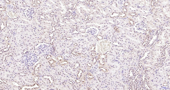
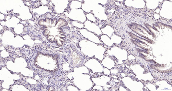
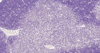
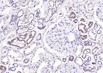
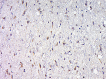
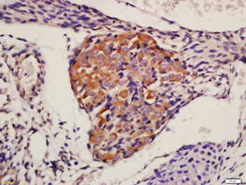
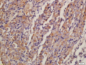
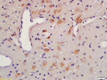
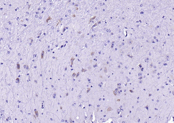
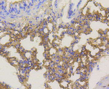
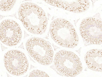
Immunohistochemistry of Rabbit Anti-AKT antibody. Tissue: (A) normal colon tissue, (B) colon tumor tissue. Fixation: formalin fixed paraffin embedded. Antigen retrieval: not required. Primary antibody: AKT antibody at 1:1000 dilution for 1 h at RT. Secondary antibody: Peroxidase rabbit secondary antibody at 1:10000 for 45 min at RT. Localization: AKT is nuclear. Staining: AKT as precipitated red signal with hematoxylin purple nuclear counterstain.
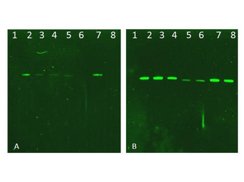
Western Blot of Rabbit AKT Antibodies. Lane 1: NIR MW protein ladder. Lane 2: AKT1, recombinant: orb346473. Lane 3: AKT1, phosphatase-treated: orb346472. Lane 4: AKT1, mutant T308A/S473A: orb346474. Lane 5: AKT2, recombinant: orb346475. Lane 6: AKT2, phosphatase-treated: orb346470. Lane 7: AKT3, recombinant: orb346476. Lane 8: AKT3, phosphatase-treated: orb346471. Load: 50 ng per lane. Blot A: orb345379 Anti-Akt pT308 used at 1:2270, Blot B: orb750474 Anti-Akt used 1:1000.
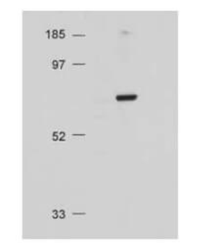
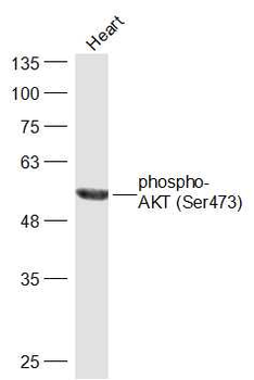
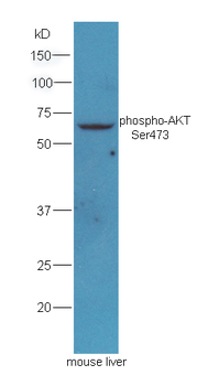
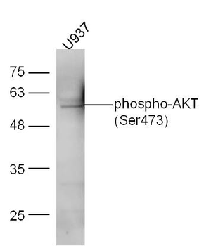
Western Blot of Rabbit Anti-AKT antibody. Lane 1: Molecular Weight. Lane 2: NIH/3T3 whole cell lysate. Load: 20 µg lysate per lane. Primary antibody: Anti-AKT antibody at 1:500 for overnight at 4°C. Secondary antibody: HRP conjugated GT-a-Rabbit IgG (orb347654) at 1:10000 preceded color development using Pierce Chemical's SuperSignal™ substrate. Block: MOPS buffer overnight at 4°C. Predicted/Observed size: 56 kDa, 56 kDa for AKT. Other band(s): none.
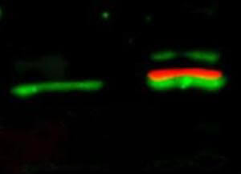
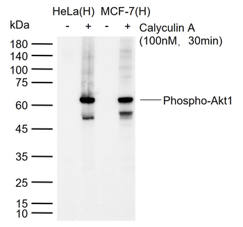
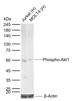
Western Blot of simultaneous detection of unphosphorylated and phosphorylated Rabbit Anti-AKT antibody. Lane 1: unstimulated NIH/3T3 lysates contain inactive unphosphorylated Akt1, green band. Lane 2: PDGF stimulated NIH/3T3 lysate contains both inactive (green band) and activated phosphorylated Akt1 (red band). Load: 35 µg per lane. Primary antibody: rabbit anti-Akt (pan) and mouse anti-Akt pS473 specific antibodies at 1:1000 for overnight at 4°C. Secondary antibody: DyLight™ 549 conjugated anti-rabbit IgG (green) and DyLight™ 649 conjugated anti-mouse IgG (red) secondary antibodies at 1:10000 for 45 min at RT. Block: 5% BLOTTO overnight at 4°C.
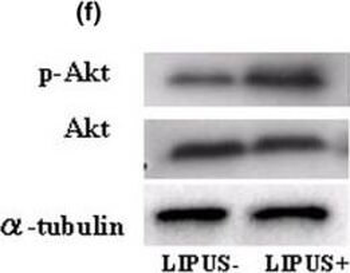
Western blotting analysis. (a) Type-II collagen. (b) Type-IX collagen. (c) Focal adhesion kinase (FAK) and phosphorylated FAK (p-FAK). (d) Paxillin and phosphorylated Paxillin (p-Paxillin). (e) Mitogen-activated protein kinase (MAPK) and phosphorylated MAPK (p-MAPK). There are no evident differences in the expression levels of total MAPK and p-MAPK between the two groups. (f) Akt and phosphorylated Akt (p-Akt). There were no differences found in the intensity the total Akt expression between the two groups, but p-Akt was found at higher levels in the LIPUS group (US+) in comparison with the control group (US-). (g) Cyclin B1 and cyclin D1. (h) Changes of proliferating cell nuclear antigen (PCNA) using MEK1 inhibitor (PD98059) and phosphatidylinositol 3-OH kinase (PI3K) inhibitor (LY294002). Chondrocytes were pretreated with MEK1 inhibitor (PD98059, 250 µm/ml) and PI3K inhibitor (LY294002, 250 µm/ml) for 12 hours and 24 hours followed by stimulation with LIPUS for 20 minutes. Each sample was harvested 2 hours after LIPUS stimulation and the influence of these inhibitors was judged in western blotting analysis of the expression of PCNA.
Documents Download
Datasheet
Product Information
Request a Document
Protocol Information
WB
Western Blot (IB, immunoblot)
IHC
Immunohistochemistry
IF
Immunofluorescence
ELISA
Enzyme-linked Immunosorbent Assay (EIA)
AKT1 Antibody (orb750475)

- 0.0
Based on 0 reviews
Participating in our Biorbyt product reviews program enables you to support fellow scientists by sharing your firsthand experience with our products.
Login to Submit a ReviewAvailable Sizes
Select a size below
Free Secondary Antibody (20 ul)0/0
Please add an antibody product to your cart first.