You have no items in your shopping cart.
Description
Research Area
Signal Transduction
Images & Validation
−Item 1 of 7
| Tested Applications | ELISA, IF, IHC, WB |
|---|---|
| Dilution Range | ELISA: 1:2,000 - 1:10,000, IHC: 1:500 - 1:2,000, IF: 1:100 - 1:1,000, WB: 1:500 - 1:2,000 |
| Reactivity | Gallus, Human, Mouse, Rat |
| Application Notes |
Key Properties
−| Antibody Type | Primary Antibody |
|---|---|
| Host | Rabbit |
| Clonality | Polyclonal |
| Isotype | Antiserum |
| Immunogen | AKT Antibody was produced from whole rabbit serum prepared by repeated immunizations with a synthetic peptide R-P-H-F-P-Q-F-S-Y-S-A-S-G-T-A corresponding to the C-terminus (460-480) of human AKT proteins conjugated to KLH using maleimide. A residue of cysteine was added to the amino terminal end to facilitate coupling. A BLAST analysis was used to suggest reactivity with this protein from rat, mouse, and chicken based on 100% homology for the immunogen sequence. |
| Target | AKT1 |
| Purity | This product was prepared from monospecific antiserum by a delipidation and defibrination. Pan Anti-AKT Antibody reacts with the AKT from human tissues. Based on sequence we expect this antibody to react as well with rat, mouse, and chicken AKT. |
| Conjugation | Unconjugated |
Storage & Handling
−| Storage | Store Anti-Akt antibody at -20° C prior to opening. Aliquot contents and freeze at -20° C or below for extended storage. Avoid cycles of freezing and thawing. Centrifuge product if not completely clear after standing at room temperature. This product is stable for several weeks at 4° C as an undiluted liquid. Dilute only prior to immediate use. |
|---|---|
| Form/Appearance | Liquid (sterile filtered) |
| Buffer/Preservatives | 0.01% (w/v) Sodium Azide |
| Concentration | 85 mg/mL |
| Expiration Date | 12 months from date of receipt. |
| Dry Ice Shipping | Please note: This product requires shipment on dry ice. A dry ice surcharge will apply. |
| Disclaimer | For research use only |
Alternative Names
−rabbit anti-AKT antibody, RAC-PK-alpha, Protein kinase B, PKB, C-AKT, RAC-alpha serine/threonine-protein kinase, Proto-oncogene c-Akt, AKT1, AKT Serine/Threonine Kinase 1, AKT 1 Antibody, AKT-1 Antibody
Similar Products
−- Item 1 of 11
Phospho-AKT (Ser473) Rabbit Polyclonal Antibody [orb11293]
WB
Bovine, Canine, Gallus, Mouse, Porcine, Rabbit, Rat, Sheep, Zebrafish
Human
Rabbit
Polyclonal
Unconjugated
50 μl, 100 μl, 200 μl - Item 1 of 9
AKT1 Antibody [orb344403]
ELISA, IF, IHC, Multiplex Assay, WB
Human, Monkey, Mouse, Rat
Mouse
Monoclonal
Unconjugated
100 μg - Item 1 of 11
Phospho-Akt (Thr450) Recombinant Rabbit Monoclonal Antibody [orb559195]
IF, IHC-Fr, IHC-P
Mouse, Rat
Human, Mouse, Rat
Rabbit
Recombinant
Unconjugated
50 μl, 100 μl, 25 μl - Item 1 of 8
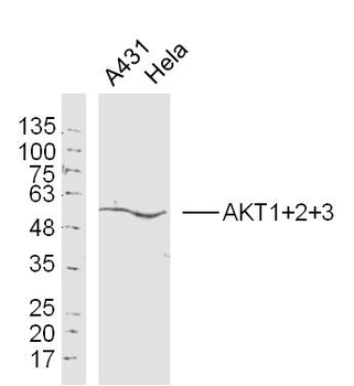
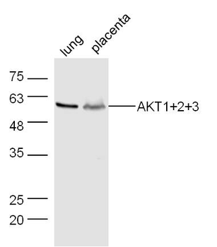
AKT1+2+3 Rabbit Polyclonal Antibody [orb155629]
FC, IF, IHC-Fr, IHC-P, WB
Bovine, Canine, Gallus, Porcine, Rabbit, Sheep
Human, Mouse, Rat
Rabbit
Polyclonal
Unconjugated
50 μl, 100 μl, 200 μl - Item 1 of 8
Phospho-Akt1 (Ser473) Recombinant Rabbit Monoclonal Antibody [orb526646]
WB
Mouse, Rat, Zebrafish
Human
Rabbit
Recombinant
Unconjugated
50 μl, 100 μl, 25 μl


Quality Guarantee
Explore bioreagents carefree to elevate your research. All our products are rigorously tested for performance. If a product does not perform as described on its datasheet, our scientific support team will provide expert troubleshooting, a prompt replacement, or a refund. For full details, please see our Terms & Conditions and Buying Guide. Contact us at [email protected].
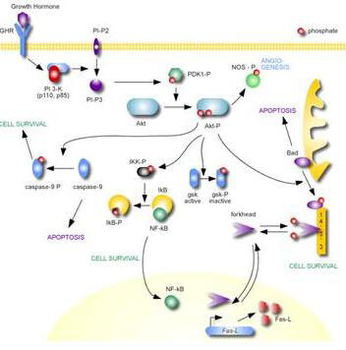
AKT Metabolic Pathway
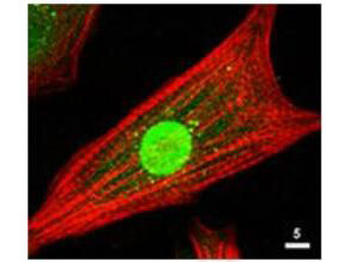
Immunofluorescence Microscopy of Rabbit Anti-AKT Antibody. Tissue: neonatal rat cardiomyocytes. Fixation: 0.5% PFA. Antigen retrieval: not required. Primary antibody: AKT antibody at 1:80 dilution for 1 h at RT. Secondary antibody: Texas-red™ conjugated rabbit secondary antibody at 1:10000 for 45 min at RT. Localization: AKT is nuclear. Staining: Anti-AKT staining appears green. Actin filaments are labeled red using a Texas-red™ conjugated phalloidin.
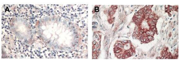
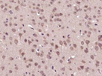
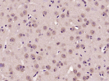
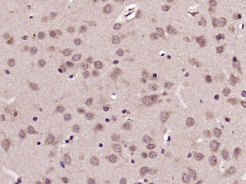
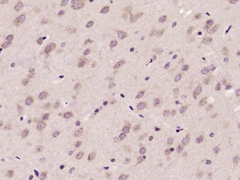
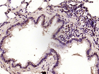
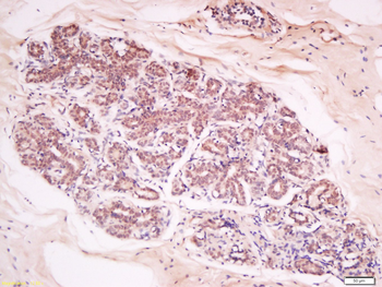
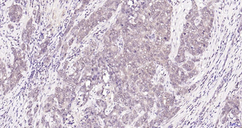
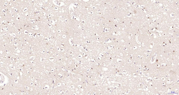
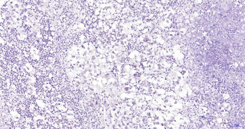
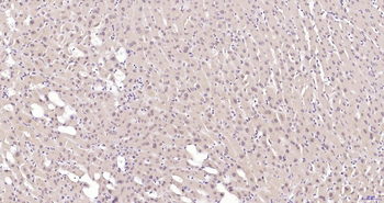
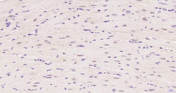
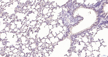
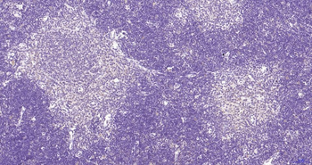
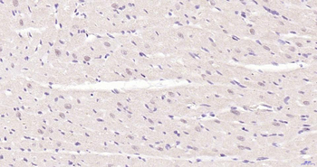
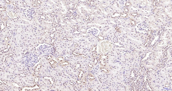
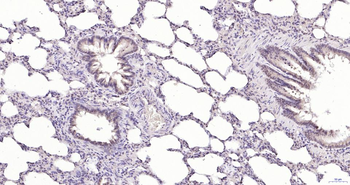
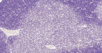
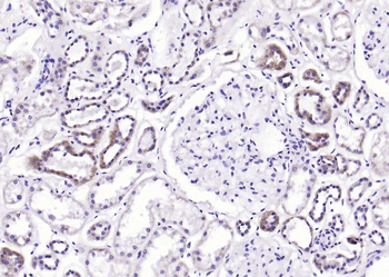
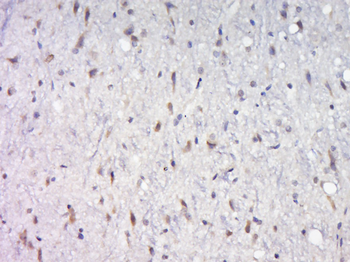
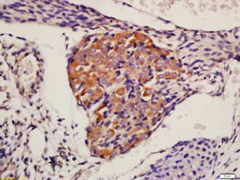
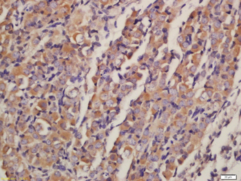
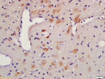
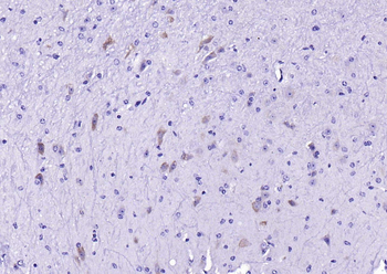
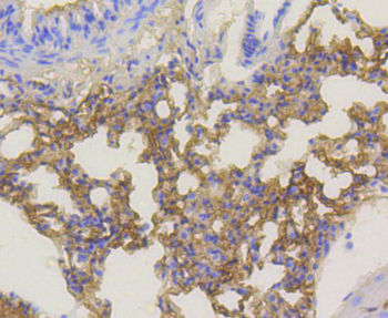
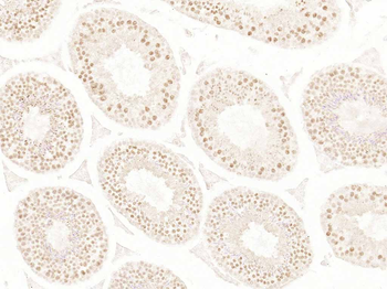
Immunohistochemistry of Rabbit Anti-AKT antibody. Tissue: (A) normal colon tissue, (B) colon tumor tissue. Fixation: formalin fixed paraffin embedded. Antigen retrieval: not required. Primary antibody: AKT antibody at 1:1000 dilution for 1 h at RT. Secondary antibody: Peroxidase rabbit secondary antibody at 1:10000 for 45 min at RT. Localization: AKT is nuclear. Staining: AKT as precipitated red signal with hematoxylin purple nuclear counterstain.
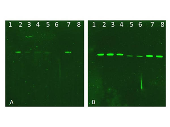
Western Blot of Rabbit AKT Antibodies. Lane 1: NIR MW protein ladder. Lane 2: AKT1, recombinant: orb346473. Lane 3: AKT1, phosphatase-treated: orb346472. Lane 4: AKT1, mutant T308A/S473A: orb346474. Lane 5: AKT2, recombinant: orb346475. Lane 6: AKT2, phosphatase-treated: orb346470. Lane 7: AKT3, recombinant: orb346476. Lane 8: AKT3, phosphatase-treated: orb346471. Load: 50 ng per lane. Blot A: orb345379 Anti-Akt pT308 used at 1:2270, Blot B: orb750474 Anti-Akt used 1:1000.
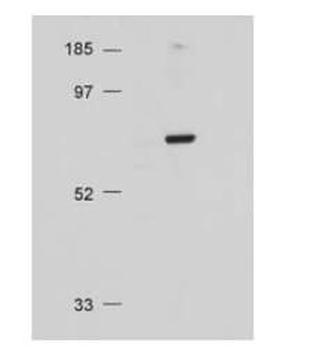
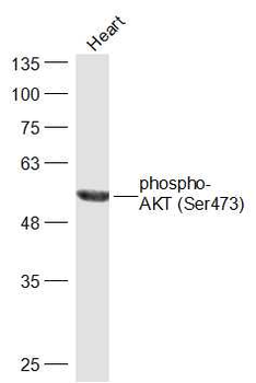
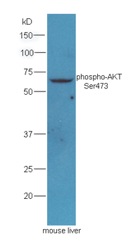
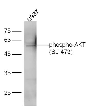
Western Blot of Rabbit Anti-AKT antibody. Lane 1: Molecular Weight. Lane 2: NIH/3T3 whole cell lysate. Load: 20 µg lysate per lane. Primary antibody: Anti-AKT antibody at 1:500 for overnight at 4°C. Secondary antibody: HRP conjugated GT-a-Rabbit IgG (orb347654) at 1:10000 preceded color development using Pierce Chemical's SuperSignal™ substrate. Block: MOPS buffer overnight at 4°C. Predicted/Observed size: 56 kDa, 56 kDa for AKT. Other band(s): none.
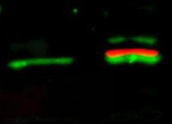
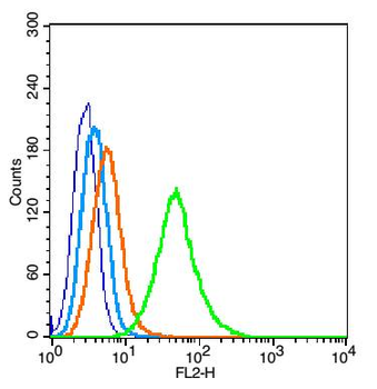
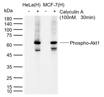
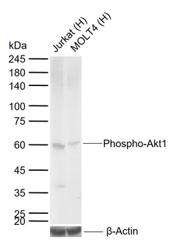
Western Blot of simultaneous detection of unphosphorylated and phosphorylated Rabbit Anti-AKT antibody. Lane 1: unstimulated NIH/3T3 lysates contain inactive unphosphorylated Akt1, green band. Lane 2: PDGF stimulated NIH/3T3 lysate contains both inactive (green band) and activated phosphorylated Akt1 (red band). Load: 35 µg per lane. Primary antibody: rabbit anti-Akt (pan) and mouse anti-Akt pS473 specific antibodies at 1:1000 for overnight at 4°C. Secondary antibody: DyLight™ 549 conjugated anti-rabbit IgG (green) and DyLight™ 649 conjugated anti-mouse IgG (red) secondary antibodies at 1:10000 for 45 min at RT. Block: 5% BLOTTO overnight at 4°C.
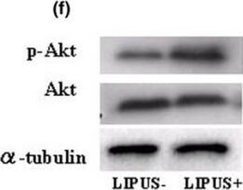
Western blotting analysis. (a) Type-II collagen. (b) Type-IX collagen. (c) Focal adhesion kinase (FAK) and phosphorylated FAK (p-FAK). (d) Paxillin and phosphorylated Paxillin (p-Paxillin). (e) Mitogen-activated protein kinase (MAPK) and phosphorylated MAPK (p-MAPK). There are no evident differences in the expression levels of total MAPK and p-MAPK between the two groups. (f) Akt and phosphorylated Akt (p-Akt). There were no differences found in the intensity the total Akt expression between the two groups, but p-Akt was found at higher levels in the LIPUS group (US+) in comparison with the control group (US-). (g) Cyclin B1 and cyclin D1. (h) Changes of proliferating cell nuclear antigen (PCNA) using MEK1 inhibitor (PD98059) and phosphatidylinositol 3-OH kinase (PI3K) inhibitor (LY294002). Chondrocytes were pretreated with MEK1 inhibitor (PD98059, 250 µm/ml) and PI3K inhibitor (LY294002, 250 µm/ml) for 12 hours and 24 hours followed by stimulation with LIPUS for 20 minutes. Each sample was harvested 2 hours after LIPUS stimulation and the influence of these inhibitors was judged in western blotting analysis of the expression of PCNA.
Documents Download
Datasheet
Product Information
Request a Document
Protocol Information
WB
Western Blot (IB, immunoblot)
IHC
Immunohistochemistry
IF
Immunofluorescence
ELISA
Enzyme-linked Immunosorbent Assay (EIA)
AKT1 Antibody (orb750474)

- 0.0
Based on 0 reviews
Participating in our Biorbyt product reviews program enables you to support fellow scientists by sharing your firsthand experience with our products.
Login to Submit a ReviewAvailable Sizes
Select a size below
Free Secondary Antibody (20 ul)0/0
Please add an antibody product to your cart first.