You have no items in your shopping cart.
Description
Research Area
Signal Transduction
Images & Validation
−Item 1 of 6
| Tested Applications | IF, IHC-P, WB |
|---|---|
| Dilution Range | WB - 1:1000, IF - 1:25, IHC-P - 1:25 |
| Reactivity | Human, Mouse |
Key Properties
−| Antibody Type | Primary Antibody |
|---|---|
| Host | Rabbit |
| Clonality | Polyclonal |
| Isotype | Rabbit IgG |
| Immunogen | This AK4 antibody is generated from a rabbit immunized with a KLH conjugated synthetic peptide between 119-153 amino acids from the Central region of human AK4. |
| Target | AK4 (HGNC:363) |
| Molecular Weight | 25268 Da |
| Conjugation | Unconjugated |
Storage & Handling
−| Storage | Maintain refrigerated at 2-8°C for up to 2 weeks. For long term storage store at -20°C in small aliquots to prevent freeze-thaw cycles |
|---|---|
| Form/Appearance | Purified polyclonal antibody supplied in PBS with 0.09% (W/V) sodium azide. This antibody is prepared by Saturated Ammonium Sulfate (SAS) precipitation followed by dialysis against PBS. |
| Expiration Date | 12 months from date of receipt. |
| Disclaimer | For research use only |
Alternative Names
−Adenylate kinase 4, mitochondrial {ECO:0000255|HAMAP-Rule:MF_03170}, AK 4 {ECO:0000255|HAMAP-Rule:MF_03170}, 27410 {ECO:0000255|HAMAP-Rule:MF_03170}, 2746 {ECO:0000255|HAMAP-Rule:MF_03170}, Adenylate kinase 3-like {ECO:0000255|HAMAP-Rule:MF_03170}, GTP:AMP phosphotransferase AK4 {ECO:0000255|HAMAP-Rule:MF_03170}, AK4 {ECO:0000255|HAMAP-Rule:MF_03170}
Quality Guarantee
Explore bioreagents carefree to elevate your research. All our products are rigorously tested for performance. If a product does not perform as described on its datasheet, our scientific support team will provide expert troubleshooting, a prompt replacement, or a refund. For full details, please see our Terms & Conditions and Buying Guide. Contact us at [email protected].
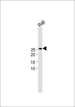
All lanes: Anti-AK4 Antibody (Center) at 1:1000 dilution + Raji whole cell lysate. Lysates/proteins at 20 µg per lane. Secondary Goat Anti-Rabbit IgG, (H+L), Peroxidase conjugated at 1/15000 dilution.Observed band size: 26kDa. Blocking/Dilution buffer: 5% NFDM/TBST.
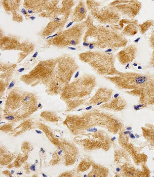
Immunohistochemical analysis of paraffin-embedded H.heart section using AK4 Antibody (Center). Diluted at 1:25 dilution. A peroxidase-conjugated goat anti-rabbit IgG at 1:400 dilution was used as the secondary antibody, followed by DAB staining.
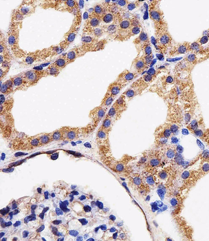
Immunohistochemical analysis of paraffin-embedded H.kidney section using AK4 Antibody (Center). Diluted at 1:25 dilution. A peroxidase-conjugated goat anti-rabbit IgG at 1:400 dilution was used as the secondary antibody, followed by DAB staining.
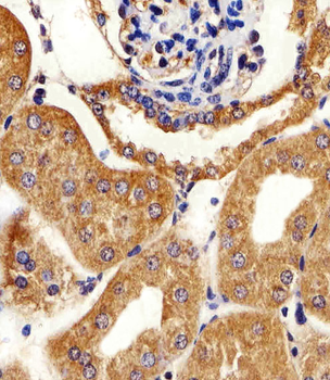
Immunohistochemical analysis of paraffin-embedded M.kidney section using AK4 Antibody (Center). Diluted at 1:25 dilution. A peroxidase-conjugated goat anti-rabbit IgG at 1:400 dilution was used as the secondary antibody, followed by DAB staining.
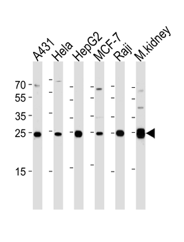
Western blot analysis of lysates from A431, Hela, HepG2, MCF-7, Raji cell line and mouse kidney tissue lysate (from left to right), using AK4 Antibody (Center). Diluted at 1:1000 at each lane. A goat anti-rabbit IgG H&L (HRP) at 1:5000 dilution was used as the secondary antibody. Lysates at 35 ug per lane.
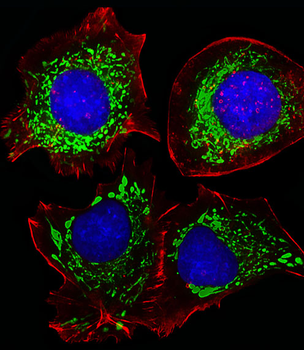
Fluorescent image of HepG2 cells stained with AK4 Antibody (Center). Diluted at 1:25 dilution. An Alexa Fluor 488-conjugated goat anti-rabbit lgG at 1:400 dilution was used as the secondary antibody (green). DAPI was used to stain the cell nuclear (blue). Cytoplasmic actin was counterstained with Alexa Fluor 555 conjugated with Phalloidin (red).
Quick Database Links
Gene Symbol
AK4 (HGNC:363)
UniProt
UniProt Details
− No UniProt data available
Documents Download
Datasheet
Product Information
Request a Document
Protocol Information
WB
Western Blot (IB, immunoblot)
IHC-P
Immunohistochemistry Paraffin
IF
Immunofluorescence
AK4 Antibody (Center) (orb1927188)

- 0.0
Based on 0 reviews
Participating in our Biorbyt product reviews program enables you to support fellow scientists by sharing your firsthand experience with our products.
Login to Submit a ReviewAvailable Sizes
Select a size below
Choose Conjugation or Carrier Free Version
Free Secondary Antibody (20 ul)0/0
Please add an antibody product to your cart first.