You have no items in your shopping cart.
Featured
KO/KD
Validated
Validated
Description
Research Area
Neuroscience
Images & Validation
−Item 1 of 15
| Tested Applications | ELISA, IF, KO/KD Validated, WB |
|---|---|
| Reactivity | Human, Mouse, Rat |
| Predicted Reactivity | Bovine |
| Application Notes |
Key Properties
−| Antibody Type | Primary Antibody |
|---|---|
| Host | Rabbit |
| Clonality | Polyclonal |
| Isotype | IgG |
| Immunogen | Anti-ADAM10 antibody (orb1239162) was raised against a peptide corresponding to 17 amino acids near the carboxy terminus of human ADAM10. The immunogen is located within the last 50 amino acids of ADAM10. |
| Target | ADAM10 |
| Molecular Weight | Predicted: 84kDObserved: 94 kD (precursor) and 68kD (mature form) |
| Purification | ADAM10 Antibody is Protein A purified. |
| Conjugation | Unconjugated |
Storage & Handling
−| Storage | Maintain refrigerated at 2-8°C for up to 2 weeks. For long term storage store at -20°C in small aliquots to prevent freeze-thaw cycles. |
|---|---|
| Form/Appearance | Liquid |
| Buffer/Preservatives | ADAM10 Antibody is supplied in PBS containing 0.02% sodium azide. |
| Concentration | 1 mg/mL |
| Expiration Date | 12 months from date of receipt. |
| Disclaimer | For research use only |
Alternative Names
−ADAM10 Antibody: RAK, kuz, AD10, AD18, MADM, CD156c, HsT18717, KUZ, Disintegrin and metalloproteinase domain-containing protein 10, CDw156, ADAM 10
Similar Products
−- Item 1 of 1
Mouse A Disintegrin and Metalloprotease 10 (ADAM10) ELISA Kit [orb778837]
Mouse
78.13-5000 pg/mL
27 pg/mL
48 T, 96 T - Item 1 of 1
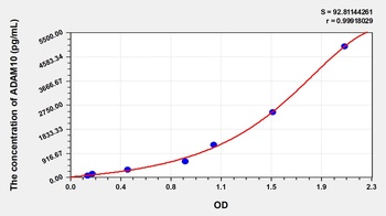
Human A Disintegrin and Metalloprotease 10 (ADAM10) ELISA Kit [orb775978]
Human
78.13-5000 pg/mL
28 pg/mL
48 T, 96 T - Item 1 of 1
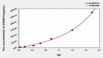
Rat A Disintegrin and Metalloprotease 10 (ADAM10) ELISA Kit [orb777008]
Rat
78.13-5000 pg/mL
27 pg/mL
48 T, 96 T - Item 1 of 1
- Item 1 of 1
Anti-ADAM10 Rabbit Polyclonal Antibody [orb2569592]
IHC, IP, WB
Human
Rabbit
Polyclonal
Unconjugated
100 μg

Quality Guarantee
Explore bioreagents carefree to elevate your research. All our products are rigorously tested for performance. If a product does not perform as described on its datasheet, our scientific support team will provide expert troubleshooting, a prompt replacement, or a refund. For full details, please see our Terms & Conditions and Buying Guide. Contact us at [email protected].
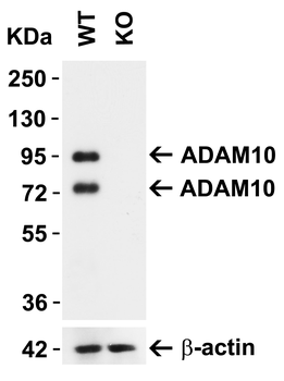
ADAM10 KO Validation in MEF Cells. Loading: 10 μg of lysate. Antibodies: ADAM10 orb1239162, 1 μg/mL and beta-actin, 1 μg/mL, 1 h incubation at RT in 5% NFDM/TBST. Secondary: Goat Anti-Rabbit IgG HRP conjugate at 1:10000 dilution. Detected both precursor ADAM10 (94KD) and mature ADAM10 (68kD).
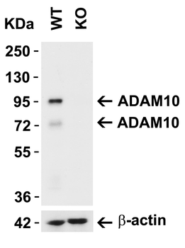
ADAM10 KO Validation in 293 Cells. Loading: 15 μg of lysate. Antibodies: ADAM10 orb1239162, 2 μg/mL and beta-actin, 1 μg/mL, 1 h incubation at RT in 5% NFDM/TBST. Secondary: Goat Anti-Rabbit IgG HRP conjugate at 1:10000 dilution. Detected both precursor ADAM10 (94KD) and mature ADAM10 (68kD).
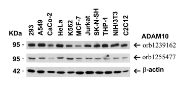
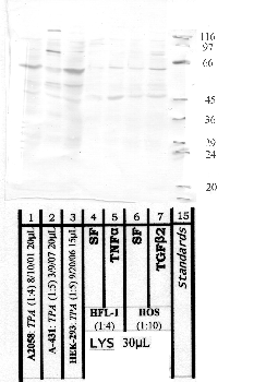
Independent Antibody Validation (IAV) via Protein Expression Profile in Human and Mouse Cell Lines. Loading: 15 μg of lysates per lane. Antibodies: ADAM10 orb1239162, 0.5 μg/mL, ADAM10 orb1255477, 1 μg/mL, and -actin orb1240312, 1 μg/mL, 1h incubation at RT in 5% NFDM/TBST. Secondary: Goat anti-rabbit IgG HRP conjugate at 1:10000 dilution.
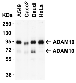
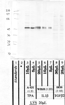
WB Validation in Human Cell Lines. Loading: 15 μg of lysate. Antibodies: ADAM10 orb1239162, 1 μg/mL, 1 h incubation at RT in 5% NFDM/TBST. Secondary: Goat Anti-Rabbit IgG HRP conjugate at 1:10000 dilution. Detected both precursor ADAM10 (94KD) and mature ADAM10 (68kD).
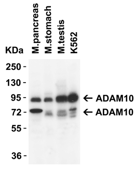
Western Blot Validation in Mouse Tissues. Loading: 15 μg of lysates per lane. Antibodies: ADAM10 orb1239162, 1 μg/mL, 1h incubation at RT in 5% NFDM/TBST. Secondary: Goat anti-rabbit IgG HRP conjugate at 1:10000 dilution. Detected both precursor ADAM10 (94KD) and mature ADAM10 (68kD).
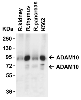
Western Blot Validation in Rat Tissues. Loading: 15 μg of lysates per lane. Antibodies: ADAM10 orb1239162, 1 μg/mL, 1h incubation at RT in 5% NFDM/TBST. Secondary: Goat anti-rabbit IgG HRP conjugate at 1:10000 dilution. Detected both precursor ADAM10 (94KD) and mature ADAM10 (68kD).
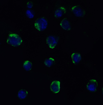
Immunofluorescence Validation of ADAM10 in MOLT4 Cells. Immunofluorescent analysis of 4% paraformaldehyde-fixed MOLT4 cells labeling ADAM10 with orb1239162 at 20 μg/mL, followed by goat anti-rabbit IgG secondary antibody at 1/500 dilution (green) and DAPI staining (blue).
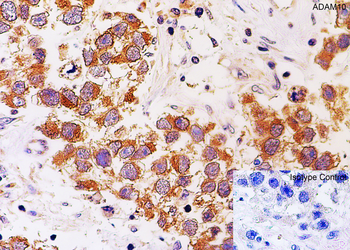
Immunohistochemistry Validation of ADAM10 in Human Testis. Immunohistochemical analysis of paraffin-embedded human testis tissue using anti-ADAM10 antibody (orb1239162) at 2 μg/ml. Tissue was fixed with formaldehyde and blocked with 10% serum for 1 h at RT; antigen retrieval was by heat mediation with a citrate buffer (pH6). Samples were incubated with primary antibody overnight at 4°C. A goat anti-rabbit IgG H&L (HRP) at 1/250 was used as secondary. Counter stained with Hematoxylin.
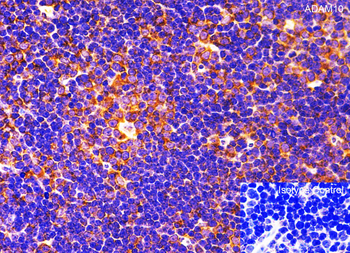
Immunohistochemistry Validation of ADAM10 in Mouse Thymus. Immunohistochemical analysis of paraffin-embedded mouse thymus tissue using anti-ADAM10 antibody (orb1239162) at 2 μg/ml. Tissue was fixed with formaldehyde and blocked with 10% serum for 1 h at RT; antigen retrieval was by heat mediation with a citrate buffer (pH6). Samples were incubated with primary antibody overnight at 4°C. A goat anti-rabbit IgG H&L (HRP) at 1/250 was used as secondary. Counter stained with Hematoxylin.
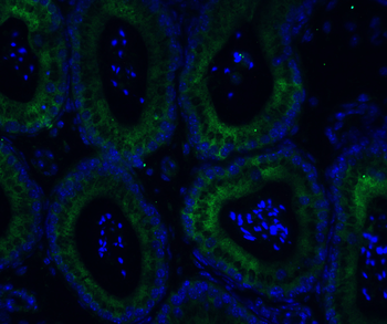
Immunofluorescence Validation of ADAM10 in Mouse Testis. Immunofluorescent analysis of 4% paraformaldehyde-fixed mouse testis labeling ADAM10 with orb1239162 at 20 μg/mL, followed by goat anti-rabbit IgG secondary antibody at 1/500 dilution (green) and DAPI staining (blue).
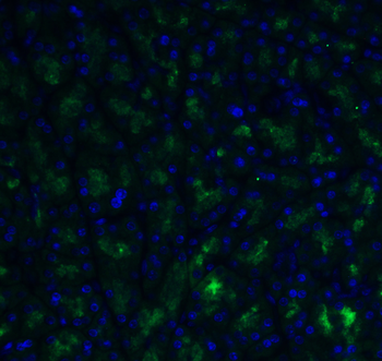
Immunofluorescence Validation of ADAM10 in Rat Testis. Immunofluorescent analysis of 4% paraformaldehyde-fixed rat testis labeling ADAM10 with orb1239162 at 20 μg/mL, followed by goat anti-rabbit IgG secondary antibody at 1/500 dilution (green) and DAPI staining (blue).
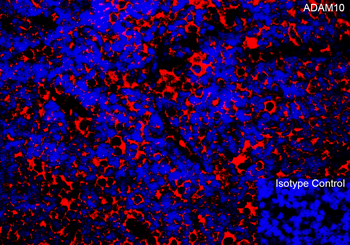
Immunofluorescence Validation of ADAM10 in Rat Thymus. Immunofluorescent analysis of 4% paraformaldehyde-fixed rat thymus labeling ADAM10 with orb1239162 at 10 μg/mL, followed by goat anti-rabbit IgG secondary antibody at 1/500 dilution (red) and DAPI staining (blue).
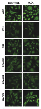
Immunofluorescence Validation of ADAM10 in primary cultures of human cerebral vascular smooth muscle cells.Detection of ADAM10 expression by anti-ADAM10 antibodies in HC-VSMC cells under control condition or in the presence of 10 μM H2O2 (oxidative stress condition) for 6 h. ADAM10 expression was not affected when exposed to oxidative stress.
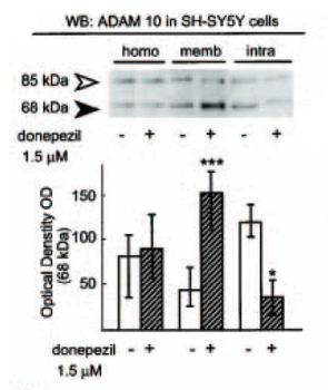
Regulated Expression Validation of ADAM10 in Human neuroblastoma. Protein expression of ADAM10 detected by anti-ADAM10 CT antibodies in control or donepezil treated SH-SY5Y cells. When treated with donepezil, the expression of mature form of ADAM10 (68kD) was up-regulated in membrane compartment as compared to the down-regulation in intracellular fractions, and was not affected in whole cell homogenate.
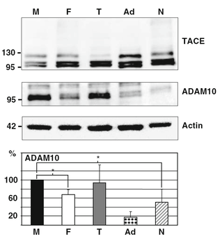
KD Validation of ADAM10 in Human embryonic kidney 293 cells overexpressing the human APP 695 isoform. Western blot analysis of ADAM10 silencing using anti-ADAM10 antibodies in HEK/APP cells. Silencing with ADAM10 siRNA (Ad) significantly decreased ADAM10 expression, and so did with Ferrochelatase siRNA (F) and N-methylprotoporphyrin IX siRNA (N), 67% and 50% reduction respectively.
Documents Download
Datasheet
Product Information
Request a Document
Protocol Information
WB
Western Blot (IB, immunoblot)
IF
Immunofluorescence
ELISA
Enzyme-linked Immunosorbent Assay (EIA)
ADAM10 Antibody (orb1239162)

- 0.0
Based on 0 reviews
Participating in our Biorbyt product reviews program enables you to support fellow scientists by sharing your firsthand experience with our products.
Login to Submit a ReviewAvailable Sizes
Select a size below
Free Secondary Antibody (20 ul)0/0
Please add an antibody product to your cart first.