You have no items in your shopping cart.
Featured
Description
Research Area
Infectious Disease & Virology
Images & Validation
−Item 1 of 11
| Tested Applications | ELISA, IF, IHC-P, WB |
|---|---|
| Reactivity | Human, Mouse, Rat |
| Predicted Reactivity | Bovine |
Key Properties
−| Antibody Type | Primary Antibody |
|---|---|
| Host | Rabbit |
| Clonality | Polyclonal |
| Isotype | IgG |
| Immunogen | Anti-ACE2 antibody (orb1239148) was raised against a peptide corresponding to 14 amino acids near the amino terminus of human ACE2. The immunogen is located within the first 50 amino acids of ACE2. |
| Target | ACE2 |
| Molecular Weight | Predicted: 93kDObserved: 130 kD (7 N-linked glycosylation) |
| Purification | ACE2 Antibody is affinity chromatography purified via peptide column. |
| Conjugation | Unconjugated |
Storage & Handling
−| Storage | Maintain refrigerated at 2-8°C for up to 2 weeks. For long term storage store at -20°C in small aliquots to prevent freeze-thaw cycles. |
|---|---|
| Form/Appearance | Liquid |
| Buffer/Preservatives | ACE2 Antibody is supplied in PBS containing 0.02% sodium azide. |
| Concentration | 1 mg/mL |
| Expiration Date | 12 months from date of receipt. |
| Disclaimer | For research use only |
Alternative Names
−ACE2 Antibody: ACEH, Angiotensin-converting enzyme 2, ACE-related carboxypeptidase, ACEH, SARS-CoV receptor, SARS-CoV-2 receptor
Similar Products
−- Item 1 of 12
ACE2 Rabbit Polyclonal Antibody [orb704533]
IF, IHC-Fr, IHC-P, WB
Mouse, Rat
Human, Mouse, Rat
Rabbit
Polyclonal
Unconjugated
50 μl, 100 μl, 200 μl - Item 1 of 11
ACE2 Recombinant Rabbit Monoclonal Antibody [orb704176]
ICC, IF, IHC-Fr, IHC-P, WB
Mouse, Rat
Human, Mouse, Rat
Rabbit
Recombinant
Unconjugated
50 μl, 100 μl, 25 μl - Item 1 of 11
ACE2 Antibody [orb1239144]
ELISA, IF, IHC-P, WB
Bovine
Human, Mouse, Rat
Rabbit
Polyclonal
Unconjugated
0.1 mg, 0.02 mg - Item 1 of 10
ACE2 Antibody [orb1239135]
ELISA, IF, IHC-P, WB
Bovine
Human, Mouse, Rat
Rabbit
Polyclonal
Unconjugated
0.1 mg, 0.02 mg - Item 1 of 9
ACE2 Antibody [orb1239146]
ELISA, IF, IHC-P, WB
Bovine
Human, Mouse, Rat
Rabbit
Polyclonal
Unconjugated
0.1 mg, 0.02 mg


Quality Guarantee
Explore bioreagents carefree to elevate your research. All our products are rigorously tested for performance. If a product does not perform as described on its datasheet, our scientific support team will provide expert troubleshooting, a prompt replacement, or a refund. For full details, please see our Terms & Conditions and Buying Guide. Contact us at [email protected].

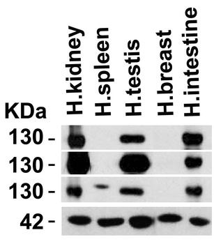
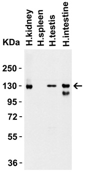
Independent Antibody Validation (IAV) via Protein Expression Profile in Human Tissues. Loading: 15 µg of lysates per lane. Antibodies: ACE2, orb1239146 (2 µg/mL), ACE2, orb1239144 (2 µg/mL), ACE2, orb1239148 (2 µg/mL) and beta-actin orb1240312 (1 µg/mL), 1h incubation at RT in 5% NFDM/TBST. Secondary: Goat anti-rabbit IgG HRP conjugate at 1:10000 dilution.
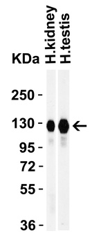
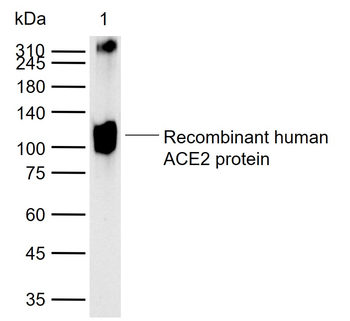
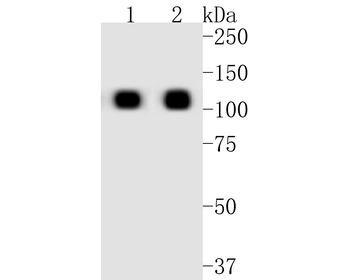
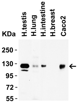
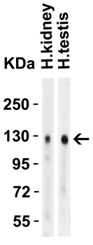
Western Blot Validation in Human Tissues. Loading: 15 µg of lysates per lane. Antibodies: ACE2, orb1239148 (1 µg/mL), 1h incubation at RT in 5% NFDM/TBST. Secondary: Goat anti-rabbit IgG HRP conjugate at 1:10000 dilution.
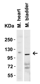
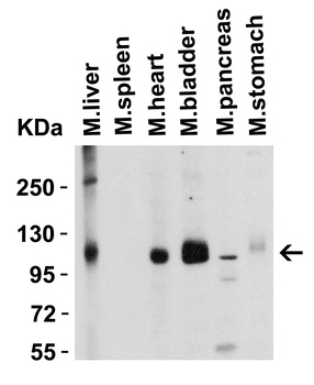
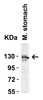
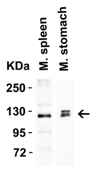
Western Blot Validation in Mouse Tissues. Loading: 15 µg of lysates per lane. Antibodies: ACE2, orb1239148 (2 µg/mL), 1h incubation at RT in 5% NFDM/TBST. Secondary: Goat anti-rabbit IgG HRP conjugate at 1:10000 dilution.
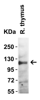
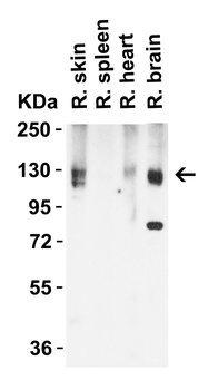
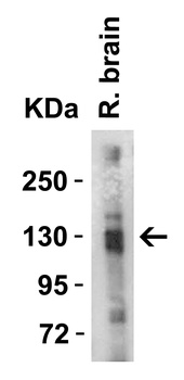
Western Blot Validation in Rat Thymus Tissue. Loading: 15 µg of lysates per lane. Antibodies: ACE2, orb1239148 (2 µg/mL), 1h incubation at RT in 5% NFDM/TBST. Secondary: Goat anti-rabbit IgG HRP conjugate at 1:10000 dilution.
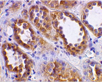
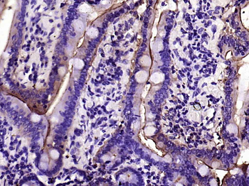
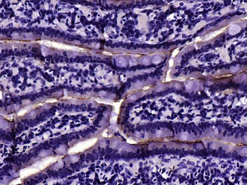
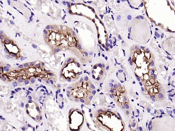
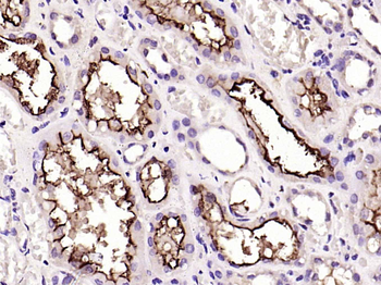
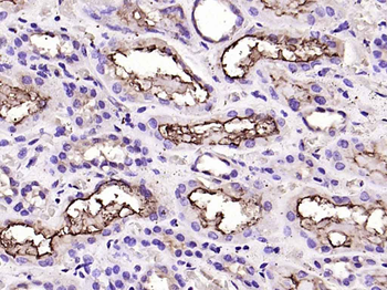
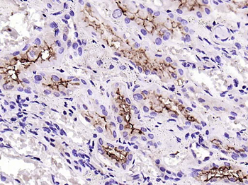
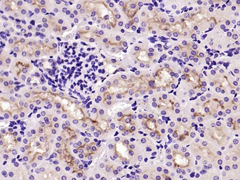
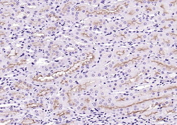
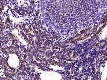
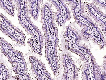
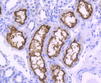
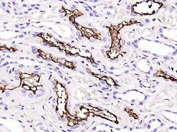
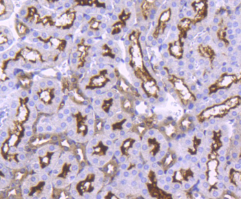
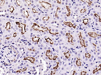
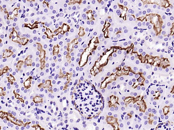
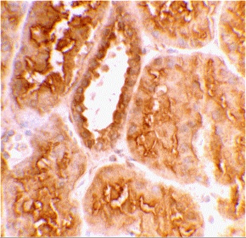
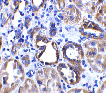
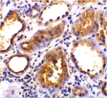
Immunohistochemistry Validation of ACE2 in Human Kidney Tissue. Immunohistochemical analysis of paraffin-embedded human kidney tissue using anti-ACE2 antibody (orb1239148) at 2 µg/ml. Tissue was fixed with formaldehyde and blocked with 10% serum for 1 h at RT; antigen retrieval was by heat mediation with a citrate buffer (pH6). Samples were incubated with primary antibody overnight at 4 °C. A goat anti-rabbit IgG H&L (HRP) at 1/250 was used as secondary. Counter stained with Hematoxylin.
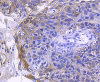
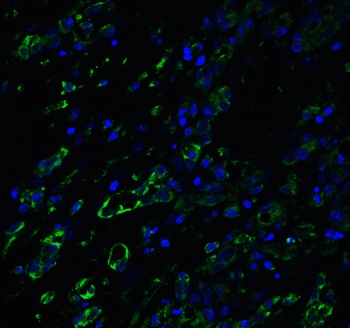
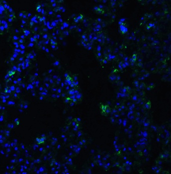
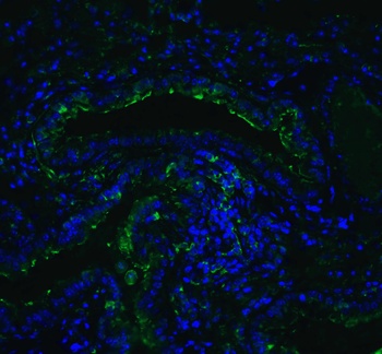
Immunofluorescence Validation of ACE2 in Human Testis Tissue. Immunofluorescent analysis of 4% paraformaldehyde-fixed human testis tissue labeling ACE-2 with orb1239148 at 20 µg/mL, followed by goat anti-rabbit IgG secondary antibody at 1/500 dilution (green) and DAPI staining (blue).
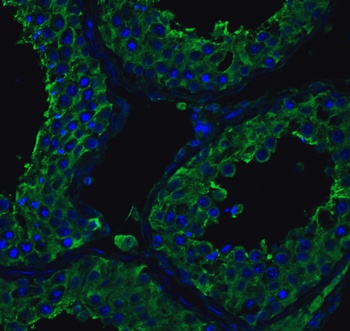
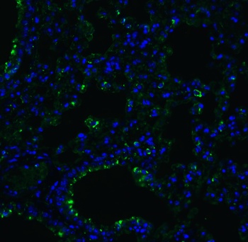
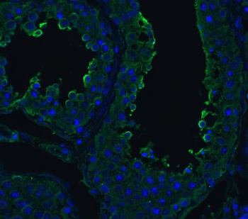
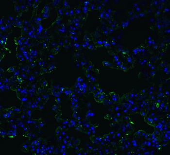
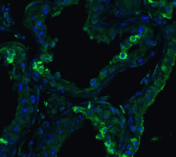
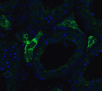
Immunofluorescence Validation of ACE2 in Human Lung Tissue. Immunofluorescent analysis of 4% paraformaldehyde-fixed human lung tissue labeling ACE-2 with orb1239148 at 20 µg/mL, followed by goat anti-rabbit IgG secondary antibody at 1/500 dilution (green) and DAPI staining (blue).
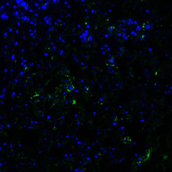
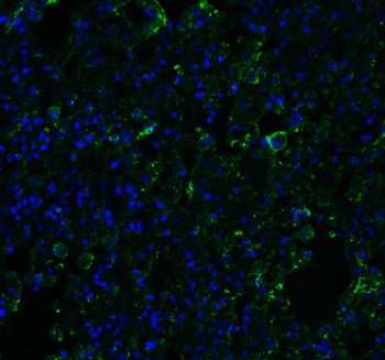
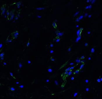
Immunofluorescence Validation of ACE2 in Mouse Lung Tissue. Immunofluorescent analysis of 4% paraformaldehyde-fixed mouse lung tissue labeling ACE-2 with orb1239148 at 20 µg/mL, followed by goat anti-rabbit IgG secondary antibody at 1/500 dilution (green) and DAPI staining (blue).
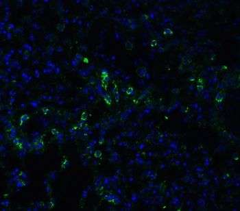
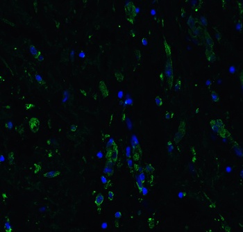
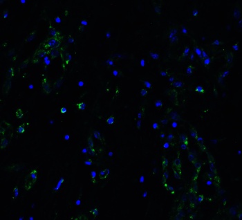
Immunofluorescence Validation of ACE2 in Rat Lung Tissue. Immunofluorescent analysis of 4% paraformaldehyde-fixed rat lung tissue labeling ACE-2 with orb1239148 at 20 µg/mL, followed by goat anti-rabbit IgG secondary antibody at 1/500 dilution (green) and DAPI staining (blue).
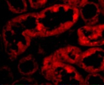
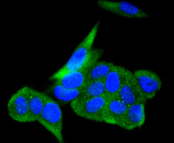
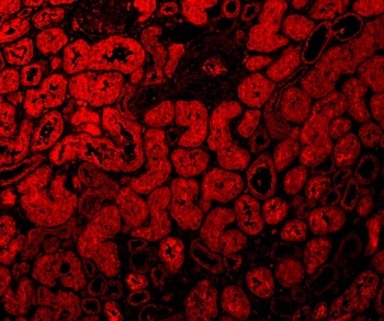
Immunofluorescence Validation of ACE2 in Human Kidney Cells. Immunofluorescent analysis of 4% paraformaldehyde-fixed human kidney cells labeling ACE2 with orb1239148 at 10 µg/mL, followed by goat anti-rabbit IgG secondary antibody at 1/500 dilution (red).
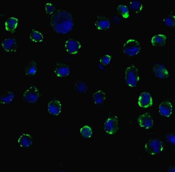
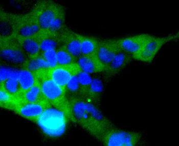
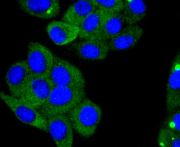
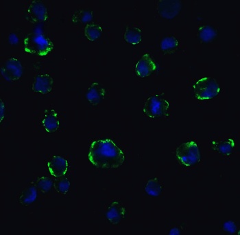
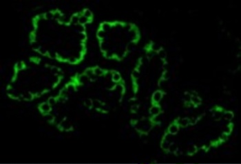
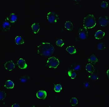
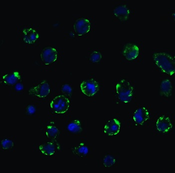
Immunofluorescence Validation of ACE2 In Caco2 Cells. Immunofluorescent analysis of 4% paraformaldehyde-fixed Caco2 cells labeling ACE2 with orb1239148 at 20 µg/mL, followed by goat anti-rabbit IgG secondary antibody at 1/500 dilution (green) and DAPI staining (blue). Image showing membrane staining on Caco2 cells.
Documents Download
Datasheet
Product Information
Request a Document
Protocol Information
WB
Western Blot (IB, immunoblot)
IHC-P
Immunohistochemistry Paraffin
IF
Immunofluorescence
ELISA
Enzyme-linked Immunosorbent Assay (EIA)
ACE2 Antibody (orb1239148)

- 0.0
Based on 0 reviews
Participating in our Biorbyt product reviews program enables you to support fellow scientists by sharing your firsthand experience with our products.
Login to Submit a ReviewAvailable Sizes
Select a size below
Free Secondary Antibody (20 ul)0/0
Please add an antibody product to your cart first.