You have no items in your shopping cart.
Description
Research Area
Immunology & Inflammation
Images & Validation
−Item 1 of 3
| Tested Applications | IHC |
|---|---|
| Dilution Range | 1:100-1:200 |
| Reactivity | Human |
| Application Notes |
Key Properties
−| Host | Rabbit |
|---|---|
| Clonality | Monoclonal |
| Isotype | IgG |
| Clone No. | MSVA-403R |
| Immunogen | Recombinant human TFE3 fragment |
| Conjugation | Unconjugated |
Storage & Handling
−| Storage | Maintain refrigerated at 2-8°C for up to 2 weeks. For long term storage store at -20°C in small aliquots to prevent freeze-thaw cycles. |
|---|---|
| Expiration Date | 12 months from date of receipt. |
| Disclaimer | For research use only |
Alternative Names
−BHLHE33
Similar Products
−- Item 1 of 3
- Item 1 of 3


Quality Guarantee
Explore bioreagents carefree to elevate your research. All our products are rigorously tested for performance. If a product does not perform as described on its datasheet, our scientific support team will provide expert troubleshooting, a prompt replacement, or a refund. For full details, please see our Terms & Conditions and Buying Guide. Contact us at [email protected].

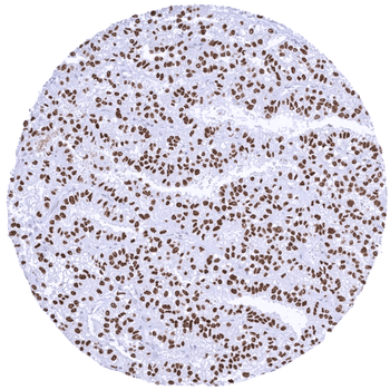
Strong nuclear TFE3 positivity in a renal cell carcinoma with TFE3 fusion.

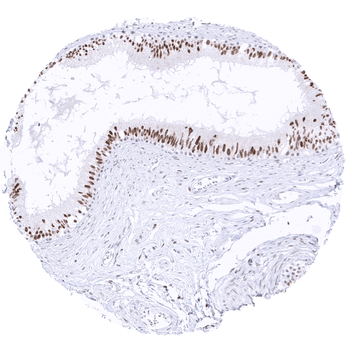
Strong TFE3 staining of epithelial cells in the corpus epididymis.

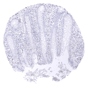
TFE3 staining is lacking in epithelial cells of the appendix.
Quick Database Links
UniProt
UniProt Details
− No UniProt data available
Documents Download
Datasheet
Product Information
Request a Document
Transcription factor E3 / TFE3 antibody (orb2276366)

- 0.0
Based on 0 reviews
Participating in our Biorbyt product reviews program enables you to support fellow scientists by sharing your firsthand experience with our products.
Login to Submit a ReviewAvailable Sizes
Select a size below
Free Secondary Antibody (20 ul)0/0
Please add an antibody product to your cart first.