You have no items in your shopping cart.
Description
Images & Validation
−Item 1 of 4
| Tested Applications | ELISA, IHC, WB |
|---|---|
| Dilution Range | WB: 1:500-1:2000 , IHC:1:20-1:200 |
| Reactivity | Human, Mouse |
Key Properties
−| Host | Rabbit |
|---|---|
| Clonality | Polyclonal |
| Isotype | IgG |
| Immunogen | Recombinant Human Thiamin pyrophosphokinase 1 protein (1-243AA) |
| Target | TPK1 |
| Purification | Antigen Affinity Purified |
| Conjugation | Unconjugated |
Storage & Handling
−| Storage | Maintain refrigerated at 2-8°C for up to 2 weeks. For long term storage store at -20°C in small aliquots to prevent freeze-thaw cycles. |
|---|---|
| Form/Appearance | Liquid |
| Buffer/Preservatives | PBS with 0.02% sodium azide, 50% glycerol, pH7.3. |
| Expiration Date | 12 months from date of receipt. |
| Disclaimer | For research use only |
Alternative Names
−hTPK1 antibody; Placental protein 20 antibody; PP20 antibody; Thiamin pyrophosphokinase 1 antibody; Thiamine diphosphokinase antibody; Thiamine kinase antibody; Thiamine pyrophosphokinase 1 antibody; THMD5 antibody; TPK1 antibody; TPK1_HUMAN antibody
Similar Products
−- Item 1 of 1
- Item 1 of 3
- Item 1 of 3
TPK1 Rabbit Polyclonal Antibody [orb632070]
ELISA, IHC, IP, WB
Human, Mouse, Rat
Rabbit
Polyclonal
Unconjugated
50 μg, 100 μg - Item 1 of 3
- Item 1 of 1
TPK1 Rabbit Polyclonal Antibody [orb2953911]
ELISA, IHC, WB
Human
Rabbit
Polyclonal
Unconjugated
50 μg, 100 μg

Quality Guarantee
Explore bioreagents carefree to elevate your research. All our products are rigorously tested for performance. If a product does not perform as described on its datasheet, our scientific support team will provide expert troubleshooting, a prompt replacement, or a refund. For full details, please see our Terms & Conditions and Buying Guide. Contact us at [email protected].

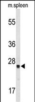
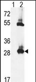
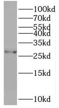
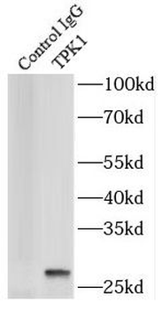
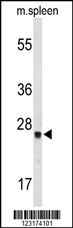
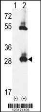
Positive WB detected in: U251 whole cell lysate(30µg), THP-1 whole cell lysate(30µg), MCF-7 whole cell lysate(30µg), K562 whole cell lysate(30µg), CT-26 whole cell lysate(30µg), PC-3 whole cell lysate(30µg), JK whole cell lysate(30µg), Mouse Liver tissue lysate(30µg), Mouse Kidneyr tissue lysate(30µg). All lanes: TPK1 antibody at 1:1000. Secondary: Goat polyclonal to rabbit IgG at 1/40000 dilution. Predicted band size: 27,14kDa, Observed band size: 27 kDa, Exposure time: 180s.

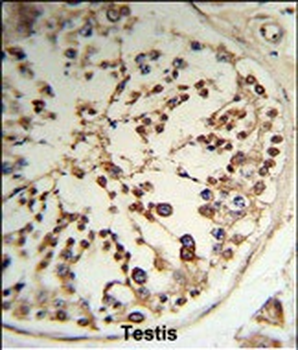
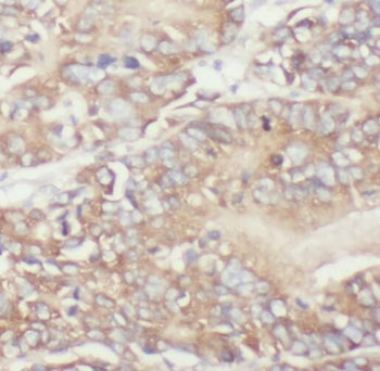
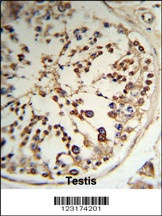
IHC image of orb41306 diluted at 1:100 and staining in paraffin-embedded human Kidney tissue performed on a Leica BondTM system. After dewaxing and hydration, antigen retrieval was mediated by high pressure in a citrate buffer (pH 6.0). Section was blocked with 10% normal goat serum 30min at RT. Then primary antibody (1% BSA) was incubated at 4°C overnight. The primary is detected by a Goat anti-rabbit polymer IgG labeled by HRP and visualized using 0.05% DAB. Secondary antibody only control: uses 1% BSA instead of primary antibody.

IHC image of orb41306 diluted at 1:100 and staining in paraffin-embedded human Heart tissue performed on a Leica BondTM system. After dewaxing and hydration, antigen retrieval was mediated by high pressure in a citrate buffer (pH 6.0). Section was blocked with 10% normal goat serum 30min at RT. Then primary antibody (1% BSA) was incubated at 4°C overnight. The primary is detected by a Goat anti-rabbit polymer IgG labeled by HRP and visualized using 0.05% DAB. Secondary antibody only control: uses 1% BSA instead of primary antibody.

IHC image of orb41306 diluted at 1:100 and staining in paraffin-embedded human breast cancer performed on a Leica BondTM system. After dewaxing and hydration, antigen retrieval was mediated by high pressure in a citrate buffer (pH 6.0). Section was blocked with 10% normal goat serum 30min at RT. Then primary antibody (1% BSA) was incubated at 4°C overnight. The primary is detected by a Goat anti-rabbit polymer IgG labeled by HRP and visualized using 0.05% DAB. Secondary antibody only control: uses 1% BSA instead of primary antibody.
Quick Database Links
Gene Symbol
TPK1
UniProt
UniProt Details
− No UniProt data available
Documents Download
Datasheet
Product Information
Request a Document
Protocol Information
WB
Western Blot (IB, immunoblot)
IHC
Immunohistochemistry
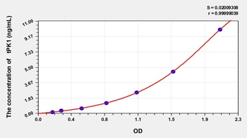
ELISA
Enzyme-linked Immunosorbent Assay (EIA)
TPK1 Antibody (orb41306)

- 0.0
Based on 0 reviews
Participating in our Biorbyt product reviews program enables you to support fellow scientists by sharing your firsthand experience with our products.
Login to Submit a ReviewAvailable Sizes
Select a size below
Choose Conjugation or Carrier Free Version
Free Secondary Antibody (20 ul)0/0
Please add an antibody product to your cart first.