You have no items in your shopping cart.
Description
Research Area
Cell Biology
Images & Validation
−Item 1 of 2
| Tested Applications | IHC, IP, WB |
|---|---|
| Dilution Range | WB: 1:100-1:1,000, IHC: 1:50-1:500, IP: 1-2 μg per 100-500 μg of total protein(1 ml of cell lysate) |
| Reactivity | Human, Mouse, Rat |
Key Properties
−| Antibody Type | Primary Antibody |
|---|---|
| Host | Mouse |
| Clonality | Polyclonal |
| Clone No. | 2G2 |
| Immunogen | Amino acids 30-301 mapping within the extracellular domain of TNF-R1 of human origin |
| Molecular Weight | Calculated MW 55 kDa |
| Purity | Affinity purified |
| Conjugation | Unconjugated |
Storage & Handling
−| Storage | Maintain refrigerated at 2-8°C for up to 2 weeks. For long term storage store at -20°C in small aliquots to prevent freeze-thaw cycles. |
|---|---|
| Form/Appearance | Liquid |
| Buffer/Preservatives | 1*TBS (pH7.4), 1%rAlbumin, 40%Glycerol. Preservative: 0.05% Sodium Azide |
| Expiration Date | 12 months from date of receipt. |
| Disclaimer | For research use only |
Similar Products
−- Item 1 of 4
TNFR1 Rabbit Polyclonal Antibody [orb100329]
FC, WB
Bovine, Canine, Equine, Porcine, Rabbit
Human, Mouse, Rat
Rabbit
Polyclonal
Unconjugated
50 μl, 200 μl, 100 μl - Item 1 of 6
TNFRSF1A Rabbit Polyclonal Antibody [orb592651]
IF, WB
Guinea pig, Mouse
Human
Rabbit
Polyclonal
Unconjugated
100 μl - Item 1 of 5
TRADD Rabbit Polyclonal Antibody [orb11504]
IF, IHC-Fr, IHC-P, WB
Rabbit
Human, Mouse, Rat
Rabbit
Polyclonal
Unconjugated
200 μl, 100 μl, 50 μl - Item 1 of 5
TRADD Antibody [orb51626]
ELISA, IF, IHC, IP, WB
Human, Rat
Rabbit
Polyclonal
Unconjugated
50 μg, 100 μg - Item 1 of 4
Human Tumor Necrosis Factor Receptor 1 (hTNFR1) Antibody [orb1562802]
ELISA, IF, IHC, WB
Human
Camelus
Monoclonal
Unconjugated
100 μg, 250 μg, 1000 μg, 2000 μg, 50 μg

Quality Guarantee
Explore bioreagents carefree to elevate your research. All our products are rigorously tested for performance. If a product does not perform as described on its datasheet, our scientific support team will provide expert troubleshooting, a prompt replacement, or a refund. For full details, please see our Terms & Conditions and Buying Guide. Contact us at [email protected].
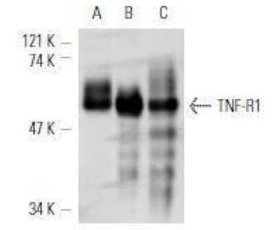
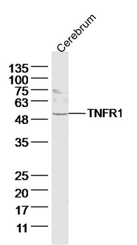
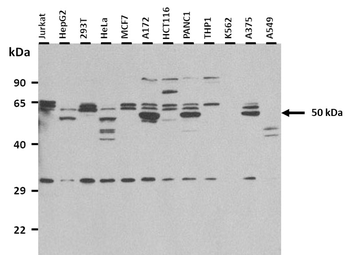
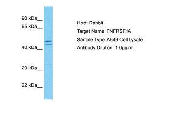
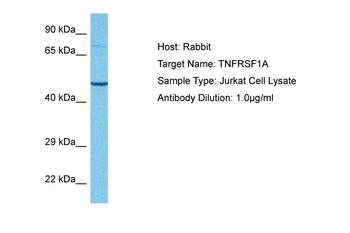
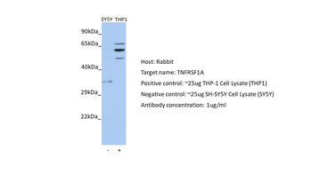
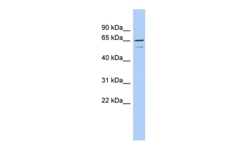
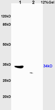
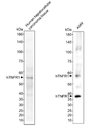
Western blot analysis of TNF-R1 expression in MCF7 (A), HeLa (B) and U-937 (C) whole cell lysates
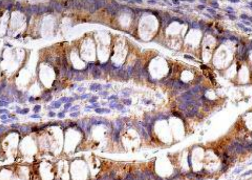
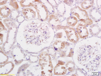
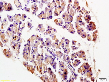
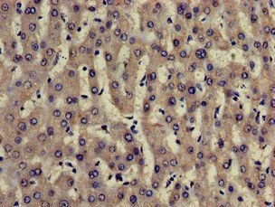
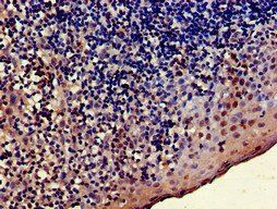
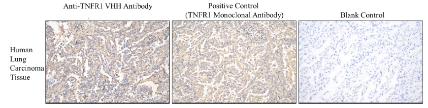
Immunoperoxidase staining of formalin fixed, paraffin-embedded human small intestine tissue showing cytoplasmic staining of glandular cells
Quick Database Links
UniProt
UniProt Details
− No UniProt data available
Documents Download
Datasheet
Product Information
Request a Document
Protocol Information
WB
Western Blot (IB, immunoblot)
IHC
Immunohistochemistry
IP
Immunoprecipitation
TNFR1 Antibody (orb621207)

- 0.0
Based on 0 reviews
Participating in our Biorbyt product reviews program enables you to support fellow scientists by sharing your firsthand experience with our products.
Login to Submit a ReviewAvailable Sizes
Select a size below
Free Secondary Antibody (20 ul)0/0
Please add an antibody product to your cart first.