You have no items in your shopping cart.
Description
Research Area
Cardiovascular Research
Images & Validation
−Item 1 of 8
| Tested Applications | IF, IHC, WB |
|---|---|
| Dilution Range | Western Blot 1:500, Immunofluorescence 1:100–1:200, Immunohistochemistry (Paraffin) 1:50–1:100 |
| Reactivity | Human, Mouse, Rat |
Key Properties
−| Antibody Type | Primary Antibody |
|---|---|
| Host | Rabbit |
| Clonality | Polyclonal |
| Isotype | Rabbit-IgG |
| Immunogen | Synthetic peptide / encompassing a sequence within the C-terminus region. |
| Target | TGF beta 1 |
| Conjugation | Unconjugated |
Storage & Handling
−| Storage | Maintain refrigerated at 2-8°C for up to 2 weeks. For long term storage store at -20°C in small aliquots to prevent freeze-thaw cycles. |
|---|---|
| Buffer/Preservatives | 100mM Tris Glycine, 1% rAlbumin, 20% Glycerol (pH7). 0.025% ProClin 300 was added as a preservative |
| Expiration Date | 12 months from date of receipt. |
| Disclaimer | For research use only |
Similar Products
−- Item 1 of 17
TGF beta 1 Rabbit Polyclonal Antibody [orb11468]
ELISA, ICC, IF, IHC-P, WB
Rabbit
Polyclonal
Unconjugated
15 μg, 200 μg, 100 μg - Item 1 of 3
TGF beta 1 Rabbit Polyclonal Antibody [orb7087]
ELISA, WB
Bovine, Canine, Guinea pig, Porcine, Rabbit, Sheep
Human, Mouse
Rabbit
Polyclonal
Unconjugated
50 μl, 100 μl, 200 μl - Item 1 of 7
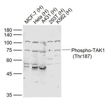
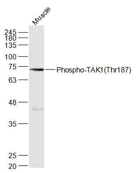
Phospho-TAK1 (Thr187) Rabbit Polyclonal Antibody [orb7049]
FC, IF, IHC-Fr, IHC-P, WB
Bovine, Equine, Gallus, Porcine, Rabbit
Human, Mouse, Rat
Rabbit
Polyclonal
Unconjugated
50 μl, 100 μl, 200 μl - Item 1 of 5
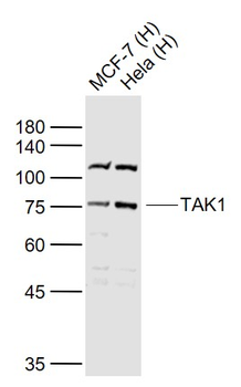
TAK1 Rabbit Polyclonal Antibody [orb7046]
FC, IF, IHC-Fr, IHC-P, WB
Bovine, Equine, Gallus, Porcine, Rabbit, Sheep
Human, Mouse, Rat
Rabbit
Polyclonal
Unconjugated
50 μl, 100 μl, 200 μl - Item 1 of 7
TGFB1 Rabbit Polyclonal Antibody [orb576457]
IF, IHC, WB
Bovine, Canine, Equine, Goat, Guinea pig, Porcine, Rat, Sheep
Human, Mouse
Rabbit
Polyclonal
Unconjugated
100 μl


Quality Guarantee
Explore bioreagents carefree to elevate your research. All our products are rigorously tested for performance. If a product does not perform as described on its datasheet, our scientific support team will provide expert troubleshooting, a prompt replacement, or a refund. For full details, please see our Terms & Conditions and Buying Guide. Contact us at [email protected].
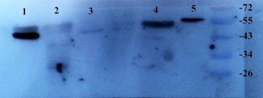
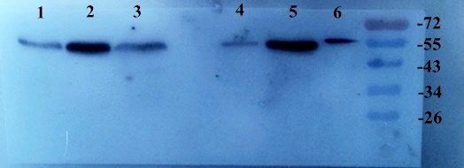
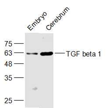
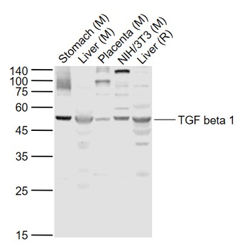
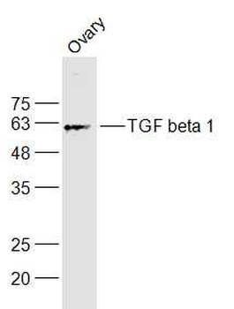
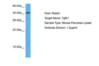
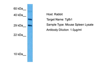
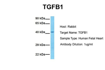
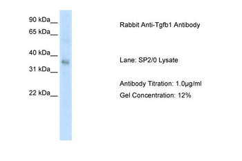
Anti TGF beta 1 antibody at 1/500 dilution. Lysates at 60 µg per lane. This blot was produced using a 12% SDS-PAGE. The gel was run at 140V for 50 minutes before being transferred onto a Nitrocellulose membrane at 18V for 60 minutes. The membrane was then blocked for an hour before being incubated with orb1294344 overnight at 4°C.
Anti TGF beta 1 antibody at 1/500 dilution. Lysates at 60 µg per lane. This blot was produced using a 12% SDS-PAGE. The gel was run at 140V for 50 minutes before being transferred onto a Nitrocellulose membrane at 18V for 60 minutes. The membrane was then blocked for an hour before being incubated with orb1294344 overnight at 4°C.
Anti TGF beta 1 antibody at 1/500 dilution. Lysates at 60 µg per lane. This blot was produced using a 12% SDS-PAGE. The gel was run at 140V for 50 minutes before being transferred onto a Nitrocellulose membrane at 18V for 60 minutes. The membrane was then blocked for an hour before being incubated with orb1294344 overnight at 4°C.
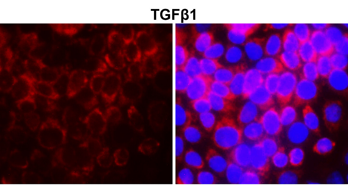
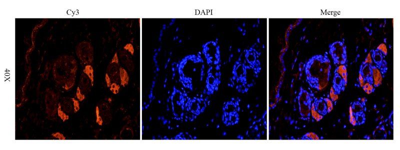
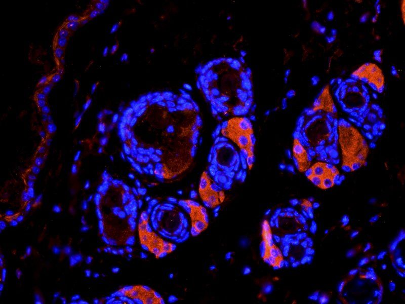
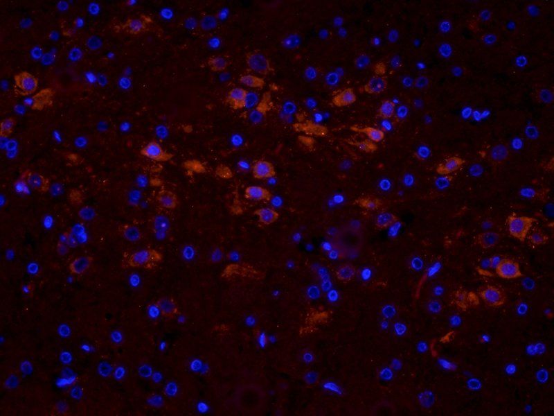
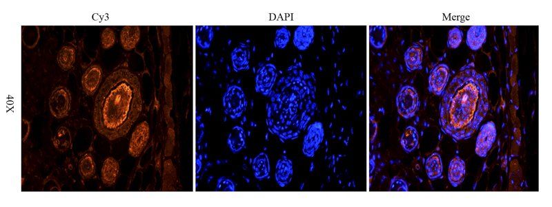
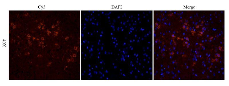
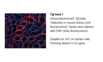
Cells were fixed with 4% paraformaldehyde for 10 min at RT, permeabilized with 0.1% NP-40 for 10 min at RT then blocked with 5% BSA for 30 min at RT. Cells were stained with orb1294344 anti-TGF beta 1 antibody (red) at 1:200 and 4°C. DAPI (blue) was used as the nuclear counter stain.
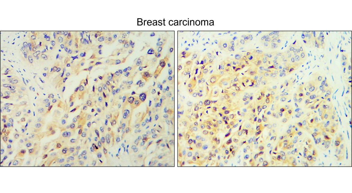
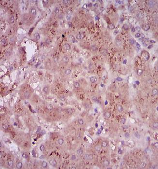
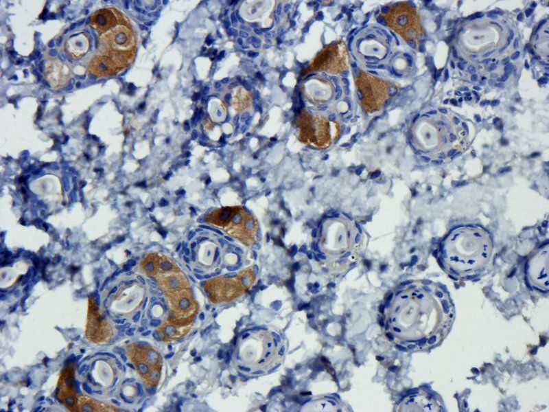
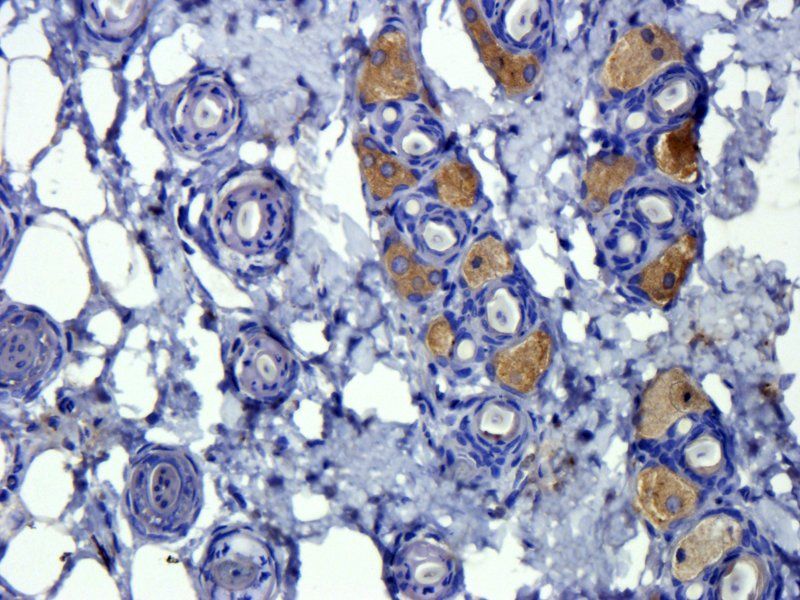
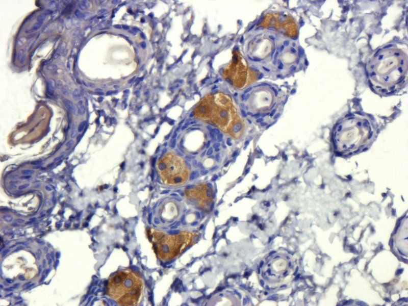
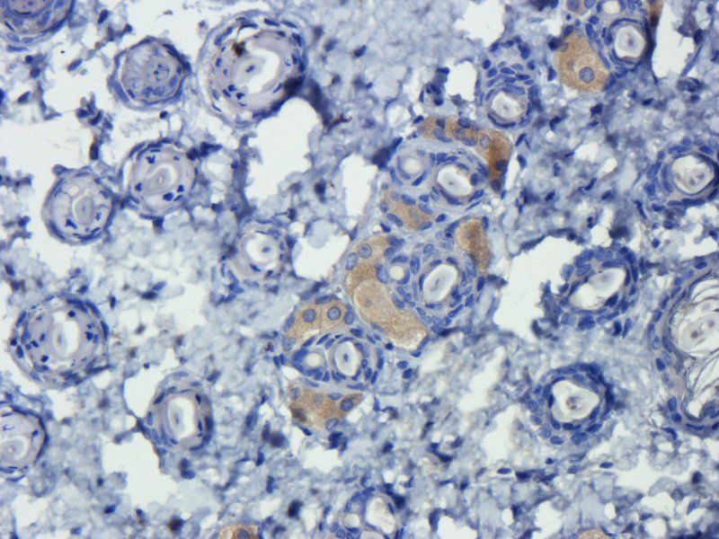
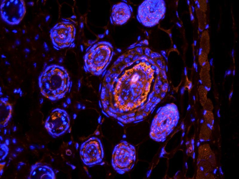
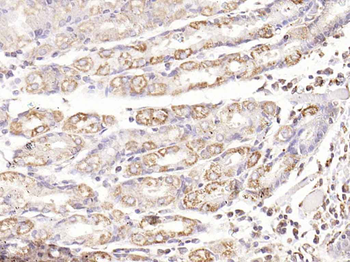
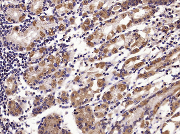
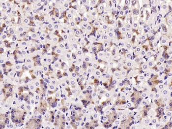
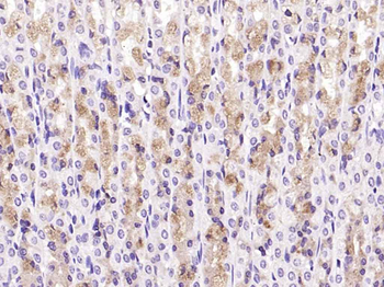
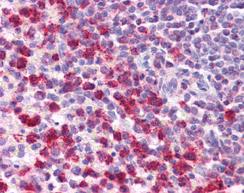
Immunohistochemical analysis of paraffin embedded Human Breast cancer tissue labeling TGF beta 1 with orb1294344 at 1/100.
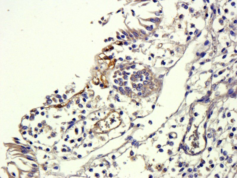
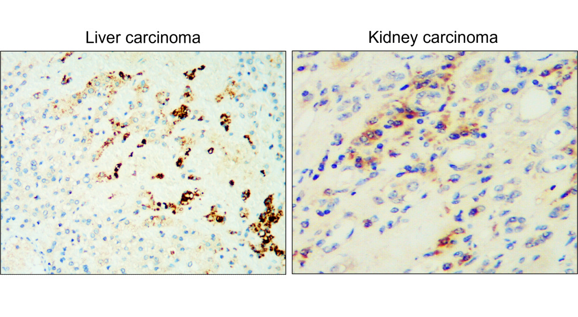
Immunohistochemical analysis of paraffin embedded Human cancer tissue labeling TGF beta 1 with orb1294344 at 1/100.
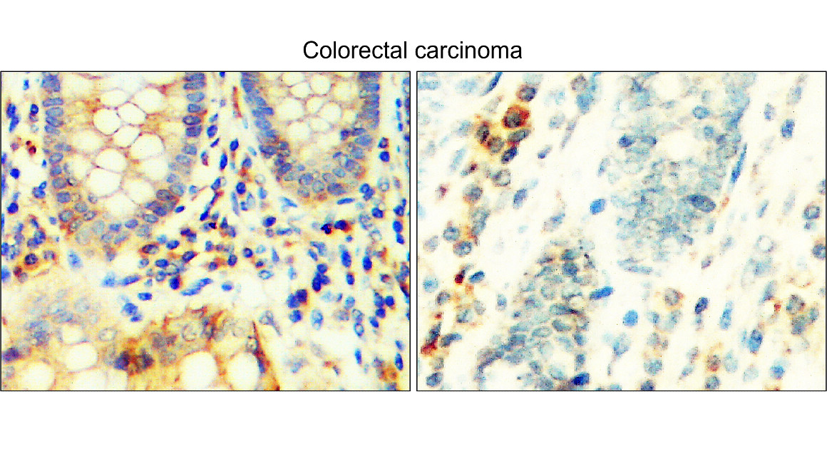
Immunohistochemical analysis of paraffin embedded Human Colorectal cancer tissue labeling TGF beta 1 with orb1294344 at 1/100.
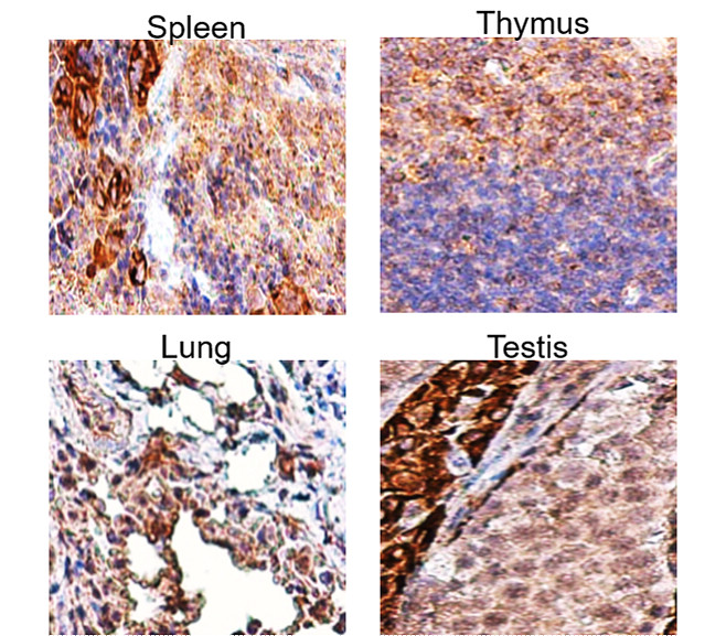
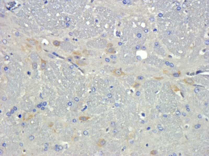
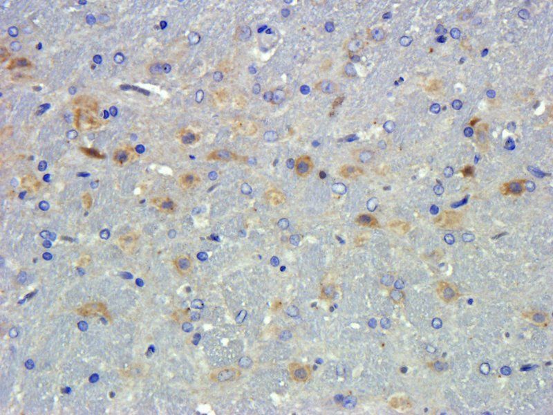
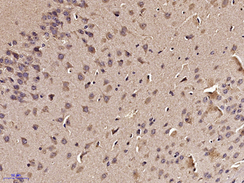
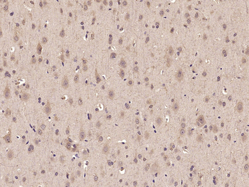
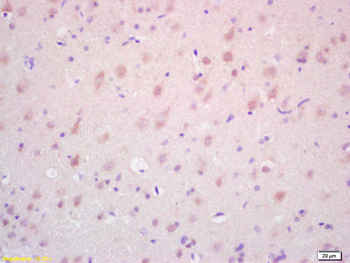
Immunohistochemical analysis of paraffin embedded mouse tissue labeling TGF beta 1 with orb1294344 at 1/100.
Quick Database Links
Gene Symbol
TGF beta 1
Documents Download
Datasheet
Product Information
Request a Document
Protocol Information
WB
Western Blot (IB, immunoblot)
IHC
Immunohistochemistry
IF
Immunofluorescence
TGF beta 1 Rabbit Polyclonal Antibody (orb1294344)

- 0.0
Based on 0 reviews
Participating in our Biorbyt product reviews program enables you to support fellow scientists by sharing your firsthand experience with our products.
Login to Submit a ReviewAvailable Sizes
Select a size below
Choose Conjugation or Carrier Free Version
Free Secondary Antibody (20 ul)0/0
Please add an antibody product to your cart first.