You have no items in your shopping cart.
Description
Images & Validation
−Item 1 of 4
| Tested Applications | ELISA, WB |
|---|---|
| Dilution Range | ELISA: 1:10,000 - 1:20,000, WB: 1:100 - 1:500 |
| Reactivity | Human |
| Application Notes |
Key Properties
−| Antibody Type | Primary Antibody |
|---|---|
| Host | Rabbit |
| Clonality | Polyclonal |
| Isotype | IgG |
| Immunogen | TGF beta 1 Antibody was prepared from whole rabbit serum produced by repeated immunizations with a synthetic peptide corresponding to amino acids near the amino terminus of the mature growth factor (112 amino acids in length). |
| Target | TGFB1 |
| Purity | Rabbit Anti-TGF beta 1 affinity purified antibody is directed against human TGFb1 protein. The product was affinity purified from monospecific antiserum by immunoaffinity chromatography. A BLAST analysis was used to suggest reactivity with this protein from most mammalian sources based on 100% homology for the immunogen sequence. Cross-reactivity is expected with TGFb1 from non-mammalian sources as only a single amino acid residue change is found within the immunogen sequence from many other organisms. |
| Conjugation | Unconjugated |
Storage & Handling
−| Storage | Store vial at -20° C prior to opening. Aliquot contents and freeze at -20° C or below for extended storage. Avoid cycles of freezing and thawing. Centrifuge product if not completely clear after standing at room temperature. This product is stable for several weeks at 4° C as an undiluted liquid. Dilute only prior to immediate use. |
|---|---|
| Form/Appearance | Liquid (sterile filtered) |
| Buffer/Preservatives | Preservative: 0.01% (w/v) Sodium Azide. Stabilizer: None; Buffer: 0.02 M Potassium Phosphate, 0.15 M Sodium Chloride, pH 7.2 |
| Concentration | 1.19 mg/ml |
| Expiration Date | 12 months from date of receipt. |
| Dry Ice Shipping | Please note: This product requires shipment on dry ice. A dry ice surcharge will apply. |
| Disclaimer | For research use only |
Alternative Names
−rabbit anti-TGFB1 antibody, rabbit anti-TGF beta 1 antibody, rabbit anti-TGF ß 1 antibody, Transforming growth factor beta-1, Latency-associated peptide, LAP, TGFB 1
Similar Products
−- Item 1 of 17
TGF beta 1 Rabbit Polyclonal Antibody [orb11468]
ELISA, ICC, IF, IHC-P, WB
Rabbit
Polyclonal
Unconjugated
15 μg, 200 μg, 100 μg - Item 1 of 3
TGF beta 1 Rabbit Polyclonal Antibody [orb7087]
ELISA, WB
Bovine, Canine, Guinea pig, Porcine, Rabbit, Sheep
Human, Mouse
Rabbit
Polyclonal
Unconjugated
50 μl, 100 μl, 200 μl - Item 1 of 9
TGF beta 1 Mouse Monoclonal Antibody [orb500906]
IF, IHC-Fr, IHC-P
Human
Mouse, Rat
Mouse
Monoclonal
Unconjugated
50 μl, 100 μl, 200 μl, 200 μg - Item 1 of 7
TGFB1 Rabbit Polyclonal Antibody [orb576457]
IF, IHC, WB
Bovine, Canine, Equine, Goat, Guinea pig, Porcine, Rat, Sheep
Human, Mouse
Rabbit
Polyclonal
Unconjugated
100 μl - Item 1 of 4
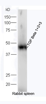
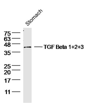
TGF Beta 1+2+3 Rabbit Polyclonal Antibody [orb7086]
FC, IF, IHC-Fr, IHC-P, WB
Bovine, Canine, Guinea pig, Porcine, Sheep
Human, Mouse, Rabbit, Rat
Rabbit
Polyclonal
Unconjugated
50 μl, 100 μl, 200 μl


Quality Guarantee
Explore bioreagents carefree to elevate your research. All our products are rigorously tested for performance. If a product does not perform as described on its datasheet, our scientific support team will provide expert troubleshooting, a prompt replacement, or a refund. For full details, please see our Terms & Conditions and Buying Guide. Contact us at [email protected].

ELISA results of purified Rabbit anti-TGF Beta 1 Antibody tested against BSA-conjugated peptide of immunizing peptide. Each well was coated in duplicate with 0.1 µg of conjugate. The starting dilution of antibody was 5 µg/ml and the X-axis represents the Log10 of a 3-fold dilution. This titration is a 4-parameter curve fit where the IC50 is defined as the titer of the antibody. Assay performed using 3% fish gel, Goat anti-Rabbit IgG Antibody Peroxidase Conjugated (Min X Bv Ch Gt GP Ham Hs Hu Ms Rt & Sh Serum Proteins) (p/n orb347654) and TMB ELISA Peroxidase Substrate (p/n orb348651).
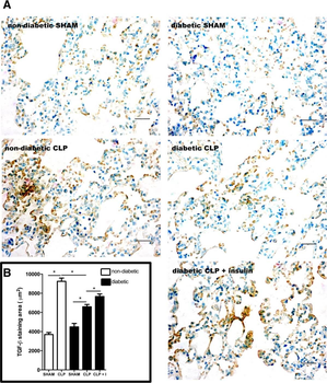
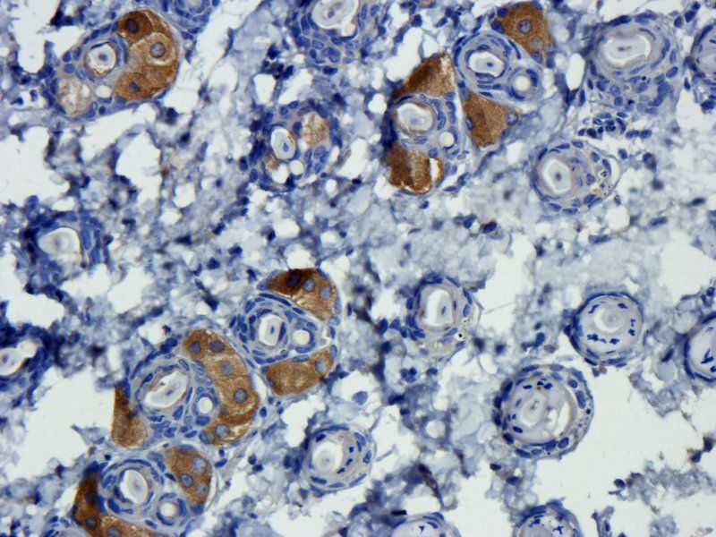
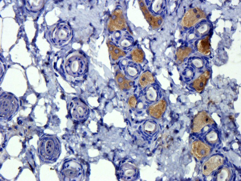
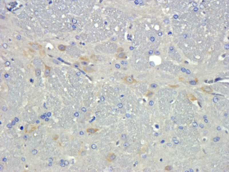
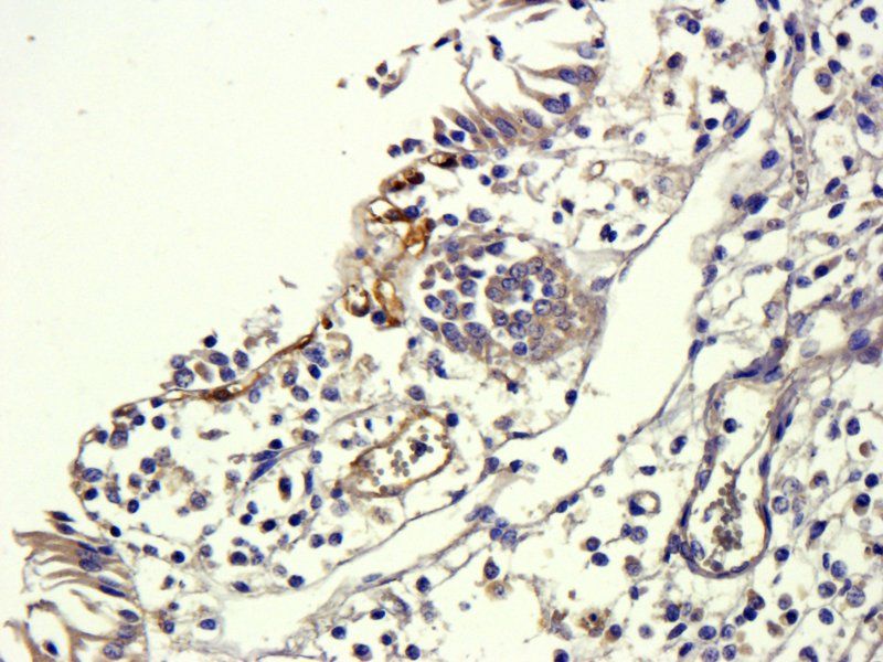
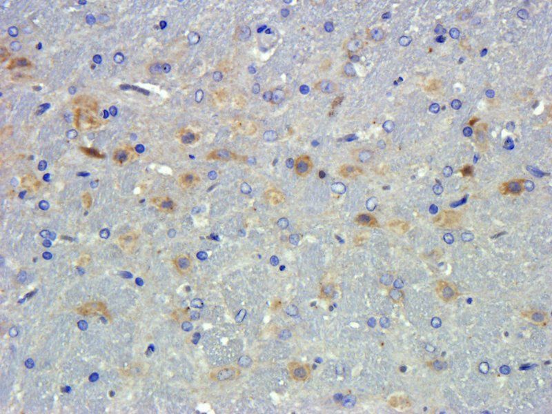
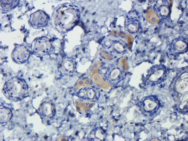
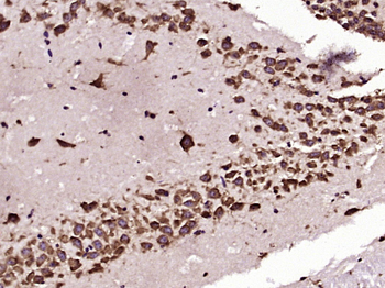
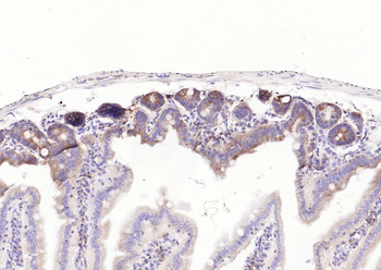
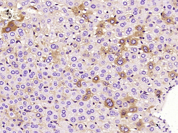
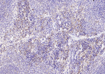
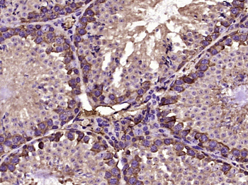
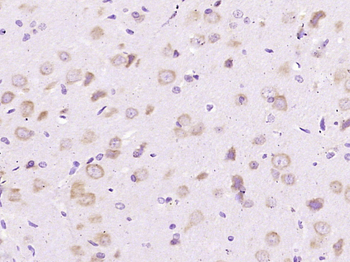
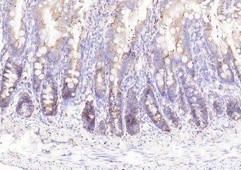
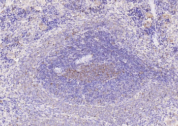
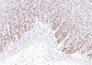
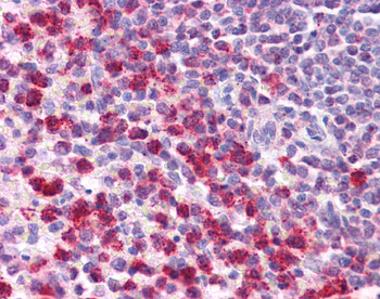
Expression of TGF-β in the lung parenchyma after CLP. Non-diabetic, diabetic and insulin-treated diabetic rats were subjected to CLP or SHAM (false operated) surgery. After 6 hours, the lungs were washed, removed and processed. The expression of TGF-β was assessed by immunohistochemistry, positive staining in brown (diffused) and nuclei in blue (A) and morphometric analysis (B) of stained area in µm2 at 400x magnification. Ten random non-coincident microscopic fields were evaluated for each group, n = 7/group. Scale bar = 20 µm. Data are presented as mean + -SEM. *p < 0.01.
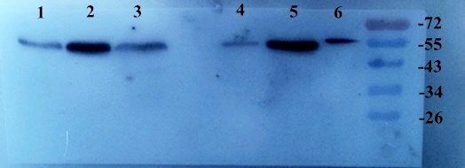
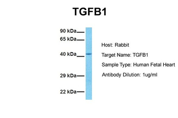
Western blot analysis using Biorbyt's Affinity Purified anti-TGF beta 1 antibody to detect human TGF beta 1. Each lane contains 250 ng of protein under non-reducing (lane 1) and reducing conditions (lane 2). Comparison to molecular weight markers (not shown) was used to estimate the indicated molecular weights. The blot was incubated with a 1:200 dilution of the antibody at room temperature for 1 h followed by detection using IRDye® 800 labeled Goat-a-Rabbit IgG (H&L) diluted 1:2500.
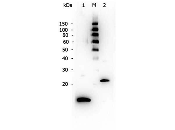
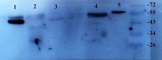
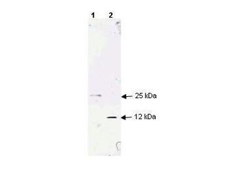
Western Blot of Rabbit anti-TGF Beta 1 antibody. Lane 1: Reduced recombinant TGF Beta 1. Lane 2: Ladder. Lane 3: Non-reduced recombinant TGF Beta 1. Load: 250 ng per lane. Primary antibody: TGF Beta 1 antibody at 1:200 for overnight at 4°C. Secondary antibody: Peroxidase rabbit secondary antibody (p/n orb347654) at 1:40000 for 30 min at RT. Block: Blocking Buffer for Fluorescent Western Blotting (orb348637). Predicted/Observed size: 12 kDa/25 kDa, 12 kDa for reduced TGF Beta 1/25 kDa for non-reduced TGF Beta 1.
Documents Download
Datasheet
Product Information
Request a Document
Protocol Information
WB
Western Blot (IB, immunoblot)
ELISA
Enzyme-linked Immunosorbent Assay (EIA)
TGFB1 Antibody (orb345420)

- 0.0
Based on 0 reviews
Participating in our Biorbyt product reviews program enables you to support fellow scientists by sharing your firsthand experience with our products.
Login to Submit a ReviewAvailable Sizes
Select a size below
Choose Conjugation or Carrier Free Version
Free Secondary Antibody (20 ul)0/0
Please add an antibody product to your cart first.