You have no items in your shopping cart.
Streptavidin Rhodamine Conjugated
SKU: orb348764
Description
Images & Validation
−Item 1 of 2
| Application Notes |
|---|
Key Properties
−| Purity | This product was prepared from pure Streptavidin as determined by electrophoresis. Assay by immunoelectrophoresis resulted in a single precipitin arc against anti-Streptavidin. |
|---|---|
| Conjugation | TRITC |
Storage & Handling
−| Storage | Store vial at 4° C prior to restoration. For extended storage aliquot contents and freeze at -20° C or below. Avoid cycles of freezing and thawing. Centrifuge product if not completely clear after standing at room temperature. This product is stable for several weeks at 4° C as an undiluted liquid. Dilute only prior to immediate use. |
|---|---|
| Form/Appearance | Lyophilized |
| Buffer/Preservatives | Preservative: 0.01% (w/v) Sodium Azide. Stabilizer: 10 mg/mL Bovine Serum Albumin (rAlbumin) - Immunoglobulin and Protease free; Buffer: 0.02 M Potassium Phosphate, 0.15 M Sodium Chloride, pH 7.2 |
| Concentration | 1.0 mg/mL |
| Expiration Date | 12 months from date of receipt. |
| Hazard Information | Non-Toxic |
| Disclaimer | For research use only |
Alternative Names
−SA, S avidin, streptococcus avidin, streptavidin TRITC
Quality Guarantee
Explore bioreagents carefree to elevate your research. All our products are rigorously tested for performance. If a product does not perform as described on its datasheet, our scientific support team will provide expert troubleshooting, a prompt replacement, or a refund. For full details, please see our Terms & Conditions and Buying Guide. Contact us at [email protected].
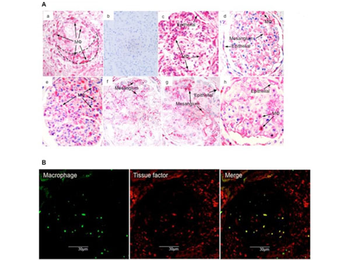
Glomerular tissue factor expression in biopsy tissues. (A) Representative tissue factor expression patterns of renal biopsy tissues as shown by immunostaining. All patients, except those with acute glomerulonephritis (AGN), had thrombinuria. (a) Crescentic glomerulonephritis (CresGN); (b) control staining of CresGN with normal mouse IgG; (c) membranoproliferative glomerulonephritis; (d) IgA nephropathy; (e) AGN; (f) minimal change glomerulopathy; (g) focal segmental glomerulosclerosis; and (h) membranous nephropathy. Mφ indicates monocytes/macrophages; Epithelial, epithelial cells; and Mesangium, mesangial areas. The broken line in (a) outlines a cellular crescent. (B) Double-staining pattern of CD68 (macrophages) and tissue factor in the CresGN glomerulus. Streptavidin Rhodamine (p/n orb348764).
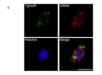
The localization of TgHoDI. C. TgHoDI-containing granular structures (false colored green), and the location of cytoplasmic mRNA granules were visualized following fluorescent in-situ hybridization using an oligo-dT-biotin probe which was subsequently revealed using rhodamine conjugated streptavidin (red) [p/n orb348764]. TgHoDI colocalizes with cytoplasmic mRNA foci (yellow). Scale bars represent 5 µm.
Documents Download
Datasheet
Product Information
Request a Document
Streptavidin Rhodamine Conjugated (orb348764)

- 0.0
Based on 0 reviews
Participating in our Biorbyt product reviews program enables you to support fellow scientists by sharing your firsthand experience with our products.
Login to Submit a ReviewAvailable Sizes
Select a size below