You have no items in your shopping cart.
Description
Research Area
Neuroscience
Images & Validation
−Item 1 of 4
| Tested Applications | IF, IHC-P, WB |
|---|---|
| Dilution Range | IF - 1:25, WB - 1:2000, IHC-P - 1:25 |
| Reactivity | Human |
Key Properties
−| Host | Mouse |
|---|---|
| Clonality | Monoclonal |
| Isotype | IgG1,k |
| Clone No. | B3578EV331X30X77 |
| Immunogen | This SMAD1 antibody is generated from a mouse immunized with a recombinant protein between 20-330 amino acids from human SMAD1. |
| Target | SMAD1 |
| Molecular Weight | 52260 Da |
| Conjugation | Unconjugated |
Storage & Handling
−| Storage | Maintain refrigerated at 2-8°C for up to 2 weeks. For long term storage store at -20°C in small aliquots to prevent freeze-thaw cycles |
|---|---|
| Form/Appearance | Purified monoclonal antibody supplied in PBS with 0.09% (W/V) sodium azide. This antibody is purified through a protein G column, followed by dialysis against PBS. |
| Expiration Date | 12 months from date of receipt. |
| Disclaimer | For research use only |
Alternative Names
−Mothers against decapentaplegic homolog 1, MAD homolog 1, Mothers against DPP homolog 1, JV4-1, Mad-related protein 1, SMAD family member 1, SMAD 1, Smad1, hSMAD1, Transforming growth factor-beta-signaling protein 1, BSP-1, SMAD1, BSP1, MADH1, MADR1
Similar Products
−- Item 1 of 7
- Item 1 of 6
SMAD1 Rabbit Polyclonal Antibody [orb500665]
IF, IHC-Fr, IHC-P, WB
Gallus
Human, Mouse, Rat
Rabbit
Polyclonal
Unconjugated
50 μl, 100 μl, 200 μl - Item 1 of 4
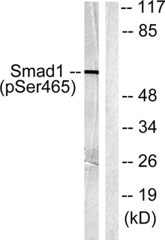
Smad1 (phospho Ser465) rabbit pAb Antibody [orb764332]
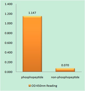
ELISA, IF, IHC, WB
Human, Mouse, Rat
Polyclonal
Unconjugated
50 μl, 100 μl - Item 1 of 5
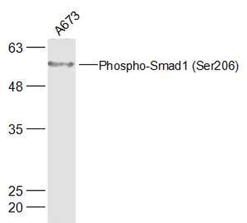
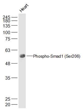
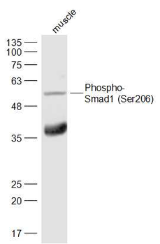
Phospho-Smad1 (Ser206) Rabbit Polyclonal Antibody [orb6976]
IF, IHC-Fr, IHC-P, WB
Rat
Human, Mouse
Rabbit
Polyclonal
Unconjugated
200 μl, 50 μl, 100 μl - Item 1 of 5
Phospho-Smad1 (Ser463) Rabbit Polyclonal Antibody [orb158426]
FC, IF, IHC-Fr, IHC-P, WB
Porcine
Human, Mouse, Rat
Rabbit
Polyclonal
Unconjugated
50 μl, 100 μl, 200 μl


Quality Guarantee
Explore bioreagents carefree to elevate your research. All our products are rigorously tested for performance. If a product does not perform as described on its datasheet, our scientific support team will provide expert troubleshooting, a prompt replacement, or a refund. For full details, please see our Terms & Conditions and Buying Guide. Contact us at [email protected].
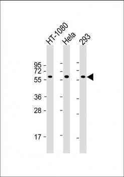
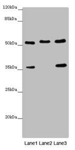
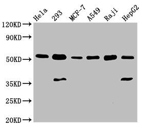
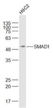
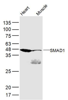
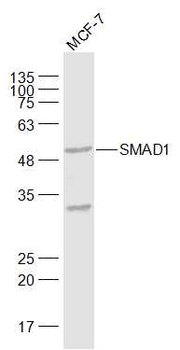
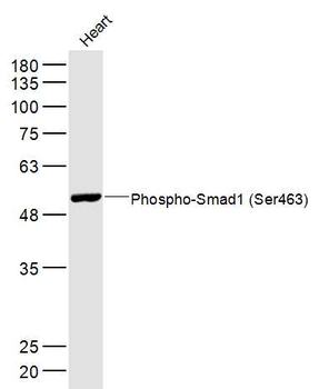
All lanes: Anti-SMAD1 Antibody at 1:2000 dilution. Lane 1: HT-1080 whole cell lysate. Lane 2: Hela whole cell lysate. Lane 3: 293 whole cell lysate. Lysates/proteins at 20 µg per lane. Secondary Goat Anti-mouse IgG, (H+L), Peroxidase conjugated at 1/10000 dilution. Predicted band size: 60 kDa. Blocking/Dilution buffer: 5% NFDM/TBST.
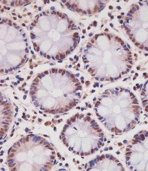
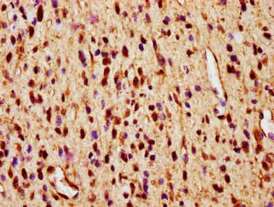
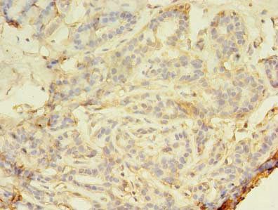
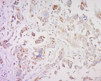
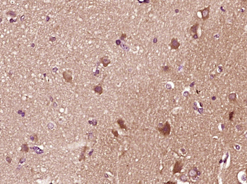
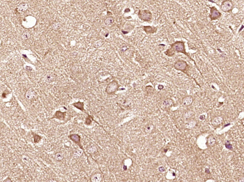
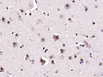
Staining SMAD1 in human colon tissue sections by Immunohistochemistry (IHC-P - paraformaldehyde-fixed, paraffin-embedded sections). Tissue was fixed with formaldehyde and blocked with 3% BSA for 0.5 hour at room temperature; antigen retrieval was by heat mediation with a citrate buffer (pH6). Samples were incubated with primary antibody (1/25) for 1 hours at 37°C. A undiluted biotinylated goat polyvalent antibody was used as the secondary antibody.
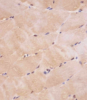
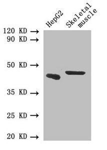
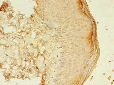
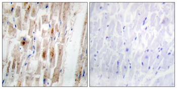
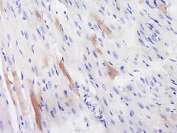
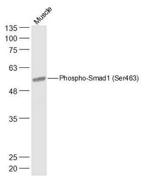
Staining SMAD1 in human skeletal muscle tissue sections by Immunohistochemistry (IHC-P - paraformaldehyde-fixed, paraffin-embedded sections). Tissue was fixed with formaldehyde and blocked with 3% BSA for 0.5 hour at room temperature; antigen retrieval was by heat mediation with a citrate buffer (pH6). Samples were incubated with primary antibody (1/25) for 1 hours at 37°C. A undiluted biotinylated goat polyvalent antibody was used as the secondary antibody.
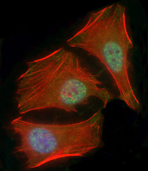
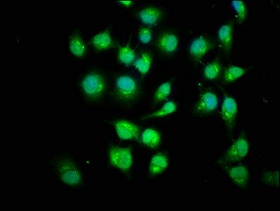
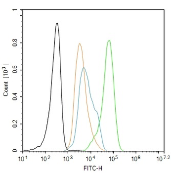
Immunofluorescent analysis of 4% paraformaldehyde-fixed, 0.1% Triton X-100 permeabilized HeLa (human cervical epithelial adenocarcinoma cell line) cells labeling SMAD1 at 1/25 dilution, followed by Dylight 488-conjugated goat anti-mouse IgG secondary antibody at 1/200 dilution (green). Immunofluorescence image showing nucleus and weak cytoplasm staining on HeLa cell line. Cytoplasmic actin is detected with Dylight 554 Phalloidin at 1/100 dilution (red). The nuclear counter stain is DAPI (blue).
Quick Database Links
Gene Symbol
SMAD1
UniProt
UniProt Details
− No UniProt data available
Documents Download
Datasheet
Product Information
Request a Document
Protocol Information
WB
Western Blot (IB, immunoblot)
IHC-P
Immunohistochemistry Paraffin
IF
Immunofluorescence
SMAD1 Antibody (orb1925575)

- 0.0
Based on 0 reviews
Participating in our Biorbyt product reviews program enables you to support fellow scientists by sharing your firsthand experience with our products.
Login to Submit a ReviewAvailable Sizes
Select a size below
Choose Conjugation or Carrier Free Version
Free Secondary Antibody (20 ul)0/0
Please add an antibody product to your cart first.