You have no items in your shopping cart.
Description
Research Area
Infectious Disease & Virology
Images & Validation
−Item 1 of 8
| Tested Applications | ELISA, IF, IHC |
|---|---|
| Reactivity | Virus |
Key Properties
−| Antibody Type | Primary Antibody |
|---|---|
| Host | Rabbit |
| Clonality | Polyclonal |
| Isotype | IgG |
| Immunogen | Anti-SARS-CoV-2 (COVID-19, 2019-nCoV) Envelope antibody (orb1239971) was raised against a peptide corresponding to 10 amino acids near the amino terminus of SARS-CoV-2 (COVID-19, 2019-nCoV) Envelope protein. The immunogen is located within the first 50 amino acids of SARS-CoV-2 (COVID-19, 2019-nCoV) Envelope. |
| Target | E |
| Purification | SARS-CoV-2 (COVID-19, 2019-nCoV) Envelope Antibody is affinity chromatography purified via peptide column. |
| Conjugation | Unconjugated |
Storage & Handling
−| Storage | Maintain refrigerated at 2-8°C for up to 2 weeks. For long term storage store at -20°C in small aliquots to prevent freeze-thaw cycles. |
|---|---|
| Form/Appearance | Liquid |
| Buffer/Preservatives | SARS-CoV-2 (COVID-19, 2019-nCoV) Envelope Antibody is supplied in PBS containing 0.02% sodium azide. |
| Concentration | 1 mg/mL |
| Expiration Date | 12 months from date of receipt. |
| Disclaimer | For research use only |
Alternative Names
−SARS-CoV-2 (COVID-19, 2019-nCoV) Envelope Antibody: Severe acute respiratory syndrome coronavirus 2 (SARS-CoV-2), Envelope protein, E protein
Similar Products
−- Item 1 of 4
SARS-CoV-2 (COVID-19) Envelope Antibody [orb1238718]
ELISA, IHC, WB
Virus
Rabbit
Polyclonal
Unconjugated
0.1 mg, 0.02 mg - Item 1 of 2
SARS-CoV-2 (COVID-19) Envelope Antibody (biotin) [orb1239041]
ELISA, IHC
Virus
Rabbit
Polyclonal
Biotin
0.1 mg, 0.02 mg - Item 1 of 2
SARS-CoV-2 (COVID-19) Envelope Antibody (HRP) [orb1239043]
ELISA, IHC
Virus
Rabbit
Polyclonal
HRP
0.1 mg, 0.02 mg 
SARS-CoV-2 (COVID-19) E protein polyclonal antibody [orb1272798]
ELISA, WB
Virus
Rabbit
Polyclonal
Unconjugated
0.1 mgSARS-CoV-2 (COVID-19) Envelope Antibody (CT) [orb1274042]
ELISA, WB
Virus
Rabbit
Polyclonal
Unconjugated
0.1 mg
Quality Guarantee
Explore bioreagents carefree to elevate your research. All our products are rigorously tested for performance. If a product does not perform as described on its datasheet, our scientific support team will provide expert troubleshooting, a prompt replacement, or a refund. For full details, please see our Terms & Conditions and Buying Guide. Contact us at [email protected].
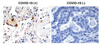
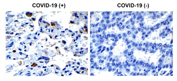
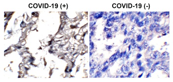
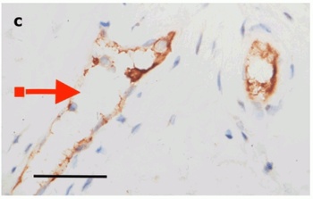
Immunohistochemistry Validation of SARS-CoV-2 (COVID-19) Envelope in COVID-19 Patient Lung. Immunohistochemical analysis of paraffin-embedded COVID-19 patient lung tissue using anti- SARS-CoV-2 (COVID-19) Envelope antibody (orb1239971, 1 µg/mL). Tissue was fixed with formaldehyde and blocked with 10% serum for 1 h at RT; antigen retrieval was by heat mediation with a citrate buffer (pH6). Samples were incubated with primary antibody overnight at 4°C. A goat anti-rabbit IgG H&L (HRP) at 1/250 was used as secondary. Counter stained with Hematoxylin. Strong signal of SARS-COV-2 envelope protein was observed in macrophage of COVID-19 patient lung, but not in non-COVID-19 patient lung.
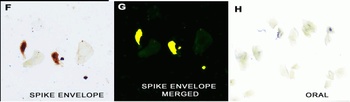
IHC/IF Validation in COVID-19 Patient Sample. (Nuovo et al., 2020) Detection of SARS-CoV-2 proteins in nasopharyngeal swab cell preparationsF-H. Co-expression of spike detected by spike antibodies (orb1239976) and envelope proteins detected by envelope antibodies (orb1239971) of SARS-CoV-2 (panel F) documented localization of each protein to glandular cells with negative squamous cells two weeks after full recovery (panel G, signal yellow). No signal was seen in oral swabs of positive cases (panel H). Both the spike and envelope protein detected by anti-spike antibodies (orb1239976, 0.2 µg/mL) and anti-envelope antibodies (orb1239971, 2 µg/mL) produced a signal in the nasopharyngeal swabs of the three cases and no signal was evident in the nasopharyngeal swabs of the seven controls.
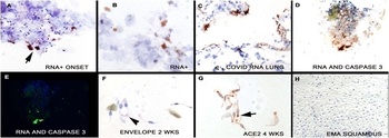
IHC Validation in COVID-19 Patient Sample. (Nuovo et al., 2020) Detection of SARS-CoV-2 Envelope protein in nasopharyngeal swab samples of COVID-19 patientsPanel F shows Envelope protein detected by envelope antibodies (orb1239971, 2 µg/mL) was still evident 2 weeks after the initial swabs (signal is red with hematoxylin counterstain), though the amount of virus was much less than at the initial swab.
IF Validation of Envelope in COVID-19 Patient Skin. (Magro et al., 2020) Detection of SARS-CoV-2 Envelope protein in the skin of COVID-19 patients that were confirmed by PCR. The skin staining shows Envelope protein expression (green) detected by envelope antibodies (orb1239971, 3 µg/mL) in mononuclear cells with hematoxylin counterstain. The staining was negative in control normal skin/lung (not shown).
IHC Validation of Envelope in COVID-19 Patient Skin. (Magro et al., 2020) Detection of SARS-CoV-2 Envelope protein in the blood vessels of COVID-19 patients that were confirmed by PCR. The staining shows Envelope protein expression (green) detected by envelope antibodies (orb1239971, 3 µg/mL) in the endothelial cytoplasms in thrombosed and normal appearing blood vessels with hematoxylin counterstain. The staining was negative in control normal skin/lung (not shown).
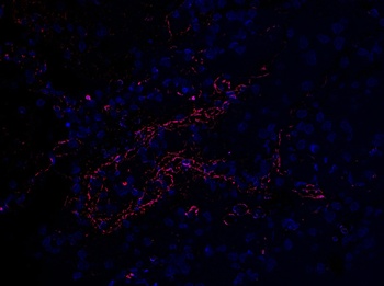
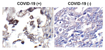
Immunofluorescence Validation of SARS-CoV-2 (COVID-19) Envelope in Human Lung Tissue from the COVID-19 Patient. Immunofluoorescent l analysis of paraffin-embedded COVID-19 patient lung tissue using anti- SARS-CoV-2 (COVID-19) Envelope antibody (orb1239971, 2 µg/mL). Tissue was fixed with formaldehyde and blocked with 10% serum for 1 h at RT; antigen retrieval was by heat mediation with a citrate buffer (pH6). Samples were incubated with primary antibody overnight at 4°C, followed by a goat anti-rabbit IgG secondary antibody at 1/500 (red) and DAPI staining (blue).
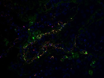
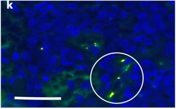
Co-expression of SARS-CoV-2 (COVID-19) Envelope and C5b-9 in Human Lung Tissue from the COVID-19 Patient. Immunofluorescent l analysis of paraffin-embedded COVID-19 patient lung tissue using anti- SARS-CoV-2 (COVID-19) Envelope antibody (orb1239971, 2 µg/mL, red) and anti-C5b-9 antibody (green). Tissue was fixed with formaldehyde and blocked with 10% serum for 1 h at RT; antigen retrieval was by heat mediation with a citrate buffer (pH6). Samples were incubated with primary antibody overnight at 4°C, followed by secondary antibodies at 1/500 and DAPI staining (blue). Co-expression was shown in yellow.
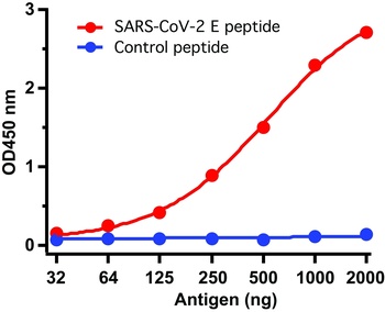
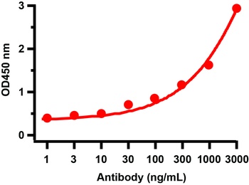
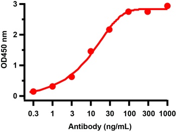
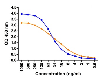
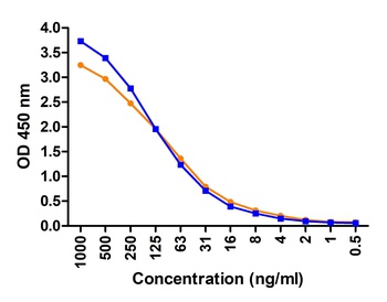
ELISA Test. Antibodies: SARS-CoV-2 (COVID-19, 2019-nCoV) Envelope Antibody, orb1239971 (1 µg/mL). A direct ELISA was performed using antigen or control peptide as coating antigen and the anti-SARS-CoV-2 (COVID-19, 2019-nCoV) Envelope antibody as the capture antibody. Secondary: Goat anti-rabbit IgG HRP conjugate at 1:20000 dilution. Detection range is from 32 ng/mL to 2000 ng/mL.
Documents Download
Datasheet
Product Information
Request a Document
Protocol Information
IHC
Immunohistochemistry
IF
Immunofluorescence
ELISA
Enzyme-linked Immunosorbent Assay (EIA)
SARS-CoV-2 (COVID-19) Envelope Antibody (orb1239971)

- 0.0
Based on 0 reviews
Participating in our Biorbyt product reviews program enables you to support fellow scientists by sharing your firsthand experience with our products.
Login to Submit a ReviewAvailable Sizes
Select a size below
Free Secondary Antibody (20 ul)0/0
Please add an antibody product to your cart first.