You have no items in your shopping cart.
Description
Research Area
Immunology & Inflammation, Metabolism Research, Neuroscience
Images & Validation
−Item 1 of 4
| Tested Applications | IHC, IP, WB |
|---|---|
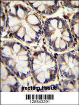
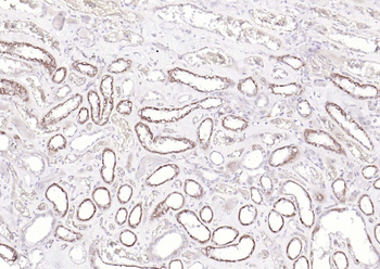
| Dilution Range | WB - 1:2,000 - 1:10,000; IP - 2 - 10 µg/mg lysate; IHC - 1:500 to 1:2,000. Epitope retrieval with citrate buffer pH6.0 is recommended for FFPE tissue sections. |
| Reactivity | Human, Mouse |
| Predicted Reactivity | Rat, Xenopus |
| Application Notes |
Key Properties
−| Antibody Type | Primary Antibody |
|---|---|
| Host | Rabbit |
| Clonality | Polyclonal |
| Isotype | IgG |
| Immunogen | Between 389 and C-terminus |
| Target | TBP1 |
| Purification | Antigen Affinity Purified |
| Conjugation | Unconjugated |
Storage & Handling
−| Storage | 2 - 8°C |
|---|---|
| Form/Appearance | Liquid |
| Buffer/Preservatives | Tris-citrate/phosphate buffer, pH 7 to 8 containing 0.09% Sodium Azide |
| Concentration | 1000 µg/ml |
| Expiration Date | 12 months from date of receipt. |
| Disclaimer | For research use only |
Alternative Names
−26S protease regulatory subunit 6A; 26S proteasome AAA-ATPase subunit RPT5; 26S proteasome regulatory subunit 6A; human immunodeficiency virus tat transactivator binding protein-1; proteasome (prosome, macropain) 26S subunit, ATPase, 3; Proteasome 26S subunit ATPase 3; proteasome subunit P50; Tat-binding protein 1; TBP1; TBP-1; testicular secretory protein Li 42
Similar Products
−- Item 1 of 4
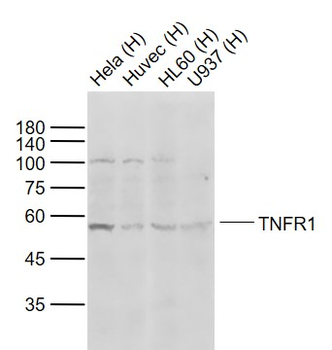
TNFR1 Rabbit Polyclonal Antibody [orb100329]
FC, WB
Bovine, Canine, Equine, Porcine, Rabbit
Human, Mouse, Rat
Rabbit
Polyclonal
Unconjugated
50 μl, 100 μl, 200 μl - Item 1 of 6
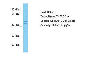
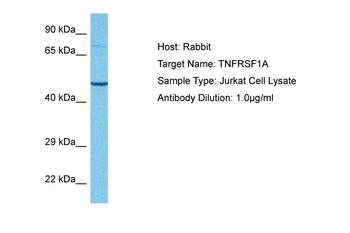
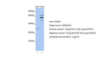
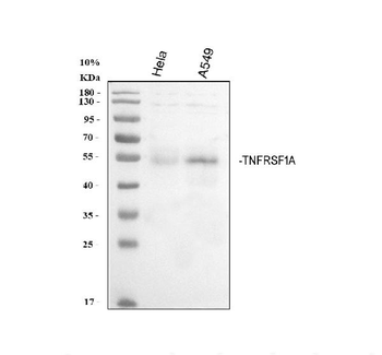
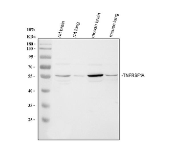
TNFRSF1A Rabbit Polyclonal Antibody [orb592651]
IF, WB
Guinea pig, Mouse
Human
Rabbit
Polyclonal
Unconjugated
100 μl - Item 1 of 4
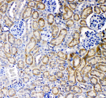
TNFRSF1A Antibody (N-term) [orb29229]
FC, IF, IHC-P, WB
Human
Rabbit
Polyclonal
Unconjugated
50 μl, 100 μl - Item 1 of 4
TNFR1 Rabbit Polyclonal Antibody [orb1062630]
IF, IHC-Fr, IHC-P, WB
Human
Rabbit
Polyclonal
Unconjugated
50 μl, 100 μl, 200 μl - Item 1 of 4
TNF Receptor I/TNFRSF1A Rabbit Polyclonal Antibody [orb443225]
ELISA, FC, IHC, WB
Human, Mouse, Rat
Rabbit
Polyclonal
Unconjugated
100 μg

Quality Guarantee
Explore bioreagents carefree to elevate your research. All our products are rigorously tested for performance. If a product does not perform as described on its datasheet, our scientific support team will provide expert troubleshooting, a prompt replacement, or a refund. For full details, please see our Terms & Conditions and Buying Guide. Contact us at [email protected].

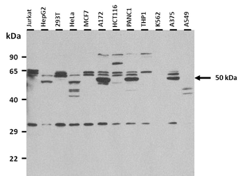
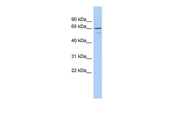
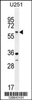
Detection of human TBP1 by western blot. Samples: Whole cell lysate (50 µg) from HEK293T, HeLa, MCF-7, Jurkat, and Hep-G2 cells prepared using NETN lysis buffer. Antibody: Affinity purified rabbit anti-TBP1 antibody (orb1524643) used for WB at 0.1 µg/ml. Detection: Chemiluminescence with an exposure time of 3 seconds.
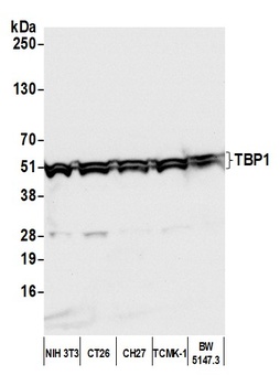
Detection of mouse TBP1 by western blot. Samples: Whole cell lysate (50 µg) from NIH 3T3, CT26, CH27, TCMK-1, and BW5147.3 cells prepared using NETN lysis buffer. Antibody: Affinity purified rabbit anti-TBP1 antibody (orb1524643) used for WB at 0.1 µg/ml. Detection: Chemiluminescence with an exposure time of 3 seconds.
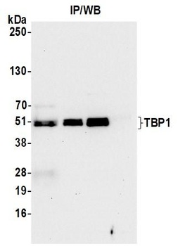
Detection of human TBP1 by western blot of immunoprecipitates. Samples: Whole cell lysate (1.0 mg per IP reaction; 10% of IP loaded) from HEK293T cells prepared using NETN lysis buffer. Antibodies: Affinity purified rabbit anti-TBP1 antibody (orb1524643) used for IP at 6 µg per reaction. TBP1 was also immunoprecipitated by a previous lot of this antibody (orb1524643) and a second antibody against a different epitope of TBP1.
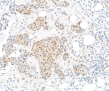
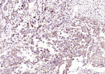
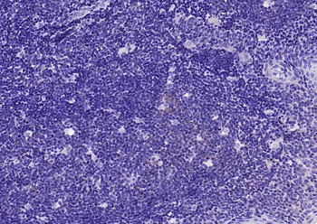
Detection of human TBP1 by immunohistochemistry.Sample: FFPE section of human breast carcinoma. Antibody: Affinity purified rabbit anti-TBP1 (orb1524643) used at a dilution of 1: 1, 000 (1µg/ml). Detection: DAB
Quick Database Links
UniProt Details
− No UniProt data available
NCBI Reference Sequences
−Associated Accession Numbers
Curated reference sequences for the gene transcript and protein product| Protein | NP_002795.2 |
|---|
Documents Download
Datasheet
Product Information
Request a Document
Protocol Information
WB
Western Blot (IB, immunoblot)
IHC
Immunohistochemistry
IP
Immunoprecipitation
Rabbit TBP1 Antibody (orb1524643)

- 0.0
Based on 0 reviews
Participating in our Biorbyt product reviews program enables you to support fellow scientists by sharing your firsthand experience with our products.
Login to Submit a ReviewAvailable Sizes
Select a size below
Choose Conjugation or Carrier Free Version
Free Secondary Antibody (20 ul)0/0
Please add an antibody product to your cart first.