You have no items in your shopping cart.
Description
Research Area
Infectious Disease & Virology
Images & Validation
−Item 1 of 2
| Tested Applications | ICC, IP, WB |
|---|---|
| Dilution Range | WB - 1:1,000 - 1:4,000; IP - 5 - 15 µg/mg lysate; ICC-IF - 1:500 - 1:2,000. Formaldehyde fixation is recommended. Permeabilization with Triton-X 100 is recommended for formaldehyde-fixed cells. |
| Reactivity | Human |
| Predicted Reactivity | Bovine, Canine, Equine, Monkey, Mouse, Rabbit, Rat |
| Application Notes |
Key Properties
−| Antibody Type | Primary Antibody |
|---|---|
| Host | Rabbit |
| Clonality | Polyclonal |
| Isotype | IgG |
| Immunogen | Between 1 and 50 |
| Target | REDD1 |
| Purification | Antigen Affinity Purified |
| Conjugation | Unconjugated |
Storage & Handling
−| Storage | 2 - 8°C |
|---|---|
| Form/Appearance | Liquid |
| Buffer/Preservatives | Tris-citrate/phosphate buffer, pH 7 to 8 containing 0.09% Sodium Azide |
| Concentration | 1000 µg/ml |
| Expiration Date | 12 months from date of receipt. |
| Disclaimer | For research use only |
Alternative Names
−Dig2; DNA damage-inducible transcript 4 protein; HIF-1 responsive protein RTP801; protein regulated in development and DNA damage response 1; REDD1; REDD-1
Similar Products
−- Item 1 of 3
DDIT4 specific Rabbit Polyclonal Antibody [orb395513]
ELISA, IHC, IP, WB
Human, Mouse, Rat
Rabbit
Polyclonal
Unconjugated
50 μg, 100 μg - Item 1 of 1
DDIT4 Rabbit Polyclonal Antibody [orb4565]
IF, IHC-Fr, IHC-P
Bovine, Equine, Human, Mouse, Porcine, Rabbit
Rat
Rabbit
Polyclonal
Unconjugated
50 μl, 100 μl, 200 μl - Item 1 of 1

- Item 1 of 1

Quality Guarantee
Explore bioreagents carefree to elevate your research. All our products are rigorously tested for performance. If a product does not perform as described on its datasheet, our scientific support team will provide expert troubleshooting, a prompt replacement, or a refund. For full details, please see our Terms & Conditions and Buying Guide. Contact us at [email protected].

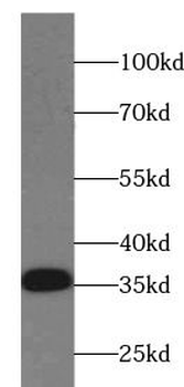
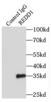
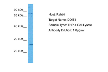
Detection of human REDD1 by western blot and immunoprecipitation. Samples: Whole cell lysate (5, 15 and 50 µg for WB; 1 mg for IP, 20% of IP loaded) from HeLa cells. Lysate was prepared from cells that had been treated with cobalt chloride (A and B) or mock treated (A). Antibodies: Affinity purified rabbit anti-REDD1 antibody orb1527090 used for WB at 0.4 µg/ml (A) and 1 µg/ml (B) and used for IP at 10 µg/mg lysate. REDD1 was also immunoprecipitated, albeit poorly, by rabbit anti-REDD1 antibody, which recognizes a downstream epitope. Detection: Chemiluminescence with exposure times of 30 seconds (A and B).
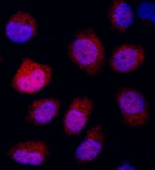
Detection of human REDD1 by immunocytochemistry.Sample: NBF-fixed asynchronous, Cobalt-treated HeLa cells. Antibody: Affinity purified rabbit anti-REDD1 (Cat. No. orb1527090) used at a dilution of 1: 500 (2 µg/ml). Detection: Red-fluorescent goat anti-rabbit IgG H&L cross-adsorbed Antibody DyLight®594 used at a dilution of 1: 100.
Quick Database Links
UniProt Details
− No UniProt data available
NCBI Reference Sequences
−Associated Accession Numbers
Curated reference sequences for the gene transcript and protein product| Protein | NP_061931.1 |
|---|
Documents Download
Datasheet
Product Information
Request a Document
Protocol Information
WB
Western Blot (IB, immunoblot)
ICC
Immunocytochemistry
IP
Immunoprecipitation
Rabbit REDD1 Antibody (orb1527090)

- 0.0
Based on 0 reviews
Participating in our Biorbyt product reviews program enables you to support fellow scientists by sharing your firsthand experience with our products.
Login to Submit a ReviewAvailable Sizes
Select a size below
Choose Conjugation or Carrier Free Version
Free Secondary Antibody (20 ul)0/0
Please add an antibody product to your cart first.