
You have no items in your shopping cart.
Electrophoresis & Western Blotting
Prestained Protein Ladders: Selection & Molecular Weight Guide
Take the guesswork out of your SDS-PAGE and Western blots. We break down how prestained protein ladders work, why apparent molecular weights shift across gels, and how to pick the right standards for your bench.
1. What Is a Prestained Protein Ladder?
A prestained protein ladder (or marker) is a mix of highly purified proteins of known molecular weights that have been covalently linked to bright, colorful dyes.
Unlike traditional unstained standards, prestained markers let you see what’s happening in real time. They act as visual landmarks on your bench, helping you track your gel run as it happens, confirm that your proteins transferred cleanly onto your nitrocellulose or PVDF membrane, and get a reliable estimate of your target protein's size on a Western blot.
Why it matters: Modern prestained ladders use multi-color reference bands (like bright green, orange, or pink indicators mixed in with standard blue bands). These make it easy to orient your gel instantly, saving you from counting tiny blue bands from the top of the cassette.
What to Expect from Standard Ladders
| Property | Typical Specification |
|---|---|
| Broad Range Markers | 10 kDa to 250 kDa (covers most standard cellular proteins) |
| High Molecular Weight | 40 kDa to 300+ kDa (best for bulky receptors, kinases, and structural proteins) |
| Low Molecular Weight | 3 kDa to 40 kDa (ideal for histones, small cytokines, and peptides) |
| Buffer Compatibility | Tris-Glycine, Bis-Tris (MES/MOPS), and Tris-Tricine |
| Storage & Handling | Store at -20°C in loading buffer (contains glycerol, SDS, and DTT/Tris) |
2. Prestained vs. Unstained Protein Markers
Both prestained and unstained markers resolve into clean size bands during SDS-PAGE, but they are built for entirely different jobs in the lab.
| Feature | Prestained Protein Ladders | Unstained Protein Ladders |
|---|---|---|
| Real-Time Gel Monitoring | Yes (track migration easily) | No (invisible until you stain the gel) |
| Membrane Transfer Verification | Yes (check transfer directly on PVDF/nitrocellulose) | No (requires Ponceau S staining first) |
| Sizing Accuracy | Good (reports apparent molecular weight) | Absolute highest accuracy |
| Mass Spectrometry Compatibility | Not recommended (dyes alter the peptides) | Ideal |
Which should you pick? Reach for prestained ladders for routine Western blotting, checking membrane transfers, and orienting gels for excision. Switch to unstained ladders when you need absolute molecular weight precision for quantitative densitometry or when prepping bands for mass spectrometry.
3. Why Do Prestained Bands Shift on Different Gels?
If you've ever noticed your marker migrating slightly differently between runs, you aren't imagining things. Prestained markers display an apparent molecular weight, not an absolute mass.
Because those colorful dye molecules are covalently bound to the proteins, they slightly change the peptide's charge-to-mass ratio and overall shape. As a result, the ladder interacts differently with various running buffers. For example, a band that migrates at 25 kDa on a standard Tris-Glycine gel might run at roughly 26 kDa in Bis-Tris MOPS—or drop to 24 kDa in Bis-Tris MES.
Bench Tip: Never assume a prestained band has the exact same molecular weight across different gel chemistries. Always check the lot-specific migration chart for your specific buffer system before sizing a critical band.
4. Recommended Prestained Protein Ladders
Biorbyt offers a dependable portfolio of ready-to-use prestained and unstained protein ladders. They're formulated for crisp band resolution, extended benchtop stability (up to 12 months), and consistent sizing across standard gel systems.
Featured Broad-Range Prestained Ladder
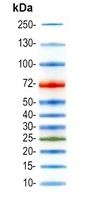
Prestained Protein Ladder (10–250 kDa) orb1289374 gel migration profile.
Featured Reagent — 10 to 250 kDa
Prestained Protein Ladder (250 kDa) — orb1289374
orb1289374 is a crisp, broad-range prestained ladder built for day-to-day SDS-PAGE monitoring and Western blot verification. Covering 10 to 180 kDa, it spans the molecular weight range of most target proteins and includes clear color-coded landmarks to keep you oriented.
MW Range: 10–250 kDa
Format: Ready-to-use liquid
Stability: Stable at -20°C
Application: SDS-PAGE & Western Blot
Complete Biorbyt Protein Ladder Portfolio
Whether you are resolving tiny peptides, massive structural proteins, or need unstained standards for exact sizing, find the right match for your workflow below:
| SKU | Profile | Product Name | MW Range |
|---|---|---|---|

|
Pre-Stained Protein Ladder Broad Range |
8–180 kDa |
|

|
Pre-Stained Protein Ladder Broad Range |
8–180 kDa |
|

|
Pre-Stained Protein Ladder Extended Broad Range |
8–250 kDa |
|
|
|
Prestained Protein Ladder Standard Broad Range |
10–180 kDa |
|

|
Pre-Stained Protein Ladder Standard Broad Range |
10–180 kDa |
|

|
Pre-Stained Protein Ladder Standard Broad Range |
10–180 kDa |
|

|
Pre-Stained Protein Ladder Standard Broad Range |
10–180 kDa |
|

|
Pre-Stained Protein Ladder Extended Broad Range |
10–250 kDa |
|

|
Unstained Protein Ladder High MW Accuracy / Low-Mid Range |
8-265KD |
|
|
|
Pre-Stained Protein Ladder Low Molecular Weight |
2.7–40 kDa |
|
|
|
Pre-Stained Protein Ladder Low Molecular Weight |
2.7–40 kDa |
Why choose an Unstained Ladder? When your primary goal is absolute molecular weight precision or downstream mass spectrometry analysis, switch to orb3169589 (8–265 kDa) or orb3169590 (2.6–70 kDa). Because these proteins don't carry bulky dye attachments, they migrate at their true, unaltered molecular weights.
5. Troubleshooting Electrophoresis Artifacts
Diffuse or Broad Bands
What causes it: You might be overloading the well, or the SDS/DTT in your sample buffer has started to break down over time.
How to fix it: Try loading just 3–5 µL per mini-gel well for Western blots (or around 10 µL if you plan to Coomassie stain)
Gel "Smiling" Effect
What causes it: Running your gel at too high a voltage generates excess heat through the center of the cassette, causing bands to curve upward at the edges.
How to fix it: Dial down the voltage (typically 100–120V is the sweet spot) and make sure your inner buffer chamber is topped up with cold running buffer.
6. Frequently Asked Questions
Should I boil a prestained protein ladder before loading?
No. Most commercial prestained ladders arrive ready to load in a buffer that has already been heat-treated and denatured. Boiling them again can strip the dyes off the proteins or cause peptide cleavage, leaving you with smeared, inaccurate bands.
Why do my ladder bands run differently on Bis-Tris gels compared to Tris-Glycine?
It comes down to buffer pH and protein charge. Those covalently attached dyes change the net charge of the proteins. Because Bis-Tris gels run at a significantly lower pH than standard Tris-Glycine gels, the ionization state of the ladder proteins shifts—changing how fast they move through the gel matrix.
How can I make my prestained ladder show up on a chemiluminescent Western blot?
While prestained dyes look great by eye, they won't inherently emit a chemiluminescent signal on film or a digital imager. To capture them alongside your target protein, you can gently mark the bands on your membrane with a chemiluminescent pen, or opt for a ladder engineered with IgG-binding domains that light up with your secondary antibody.