You have no items in your shopping cart.
On Promotion
Category
Tested Applications
WB 42960ELISA 22433SDS-PAGE 15106Blocking 11025IHC 2122MS 2019HPLC 619LF 518CLIA 507EIA 383Cell Culture 288FA 198IP 98IF 88ChIP 57DOT 49IA 38Functional Studies 24FC 21In vitro 15In vivo 14ATPase Activity Assay 9Control 8CBA 6ICC 6Purification 6FACS 2GICA 2IAC 2IB 2ICA 2PCR 2SBR 22D-PAGE 1CoIP 1DARTs Assay 1Double Diffusion 1ELISpot 1FLISA 1HAI 1ID 1IEP 1RIA 1SB 1Turbidimetry 1cDNA Synthesis 1
Reactivity
Human 16895Mouse 6092Rat 4647Monkey 1687Bovine 1675Porcine 1084Canine 675Gallus 657Rabbit 425Virus 392Zebrafish 387Sheep 297Other 56Bacteria 53Hamster 10Goat 9Plant 9Protozoa 9Feline 8Guinea pig 7Parasite 7Equine 5Camelus 3E. coli 3Fungi 2H. pylori 2Primate 2Rat and Mouse 2A. thaliana 1Algae 1Aspergillus 1Ferret 1HCMV 1HSV-1 1Jellyfish 1Marmoset 1Rabies virus 1Yeast 1
Predicted Reactivity
Conjugation
Biologically Active
Featured Product
All Products
- Featured
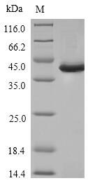
 Item 1 of 3Human SPI1 protein [orb419162]Featured
Greater than 85% as determined by SDS-PAGE.
35.1 kDa
E.coli
20 μg, 100 μg, 1 mg - Item 1 of 4
SDS-PAGE
Unconjugated
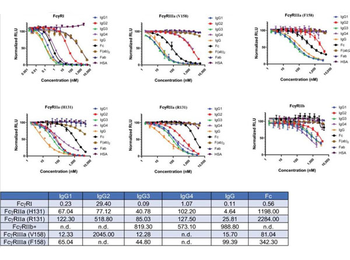
Human IgG Fab fragment was prepared from normal serum by a multi-step process which includes delipidation, salt fractionation and ion exchange chromatography followed by papain digestion and extensive dialysis against the buffer stated above. Human IgG Fab fragment assayed by immunoelectrophoresis resulted in a single precipitin arc against anti-Human Serum, anti- Human IgG and anti- Human IgG F(ab’)2. No reaction was observed against anti- Human IgG F(c) or anti-Papain.
Human
2 mg - Item 1 of 2
- FeaturedItem 1 of 3Featured
Unconjugated
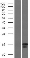
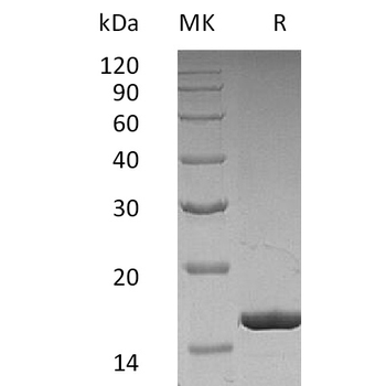
Greater than 95% as determined by reducing SDS-PAGE.
16.6 KDa
Mammalian
10 μg, 50 μg - FeaturedItem 1 of 4Featured
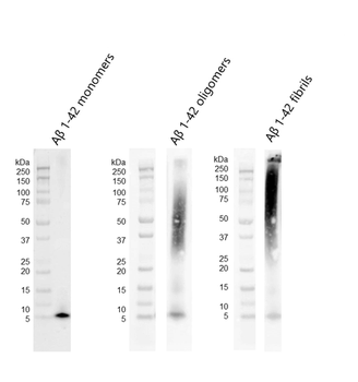
In vitro, In vivo, WB
>98%
4.5 kDa
Synthetic
100 μg - FeaturedItem 1 of 3Featured
Unconjugated
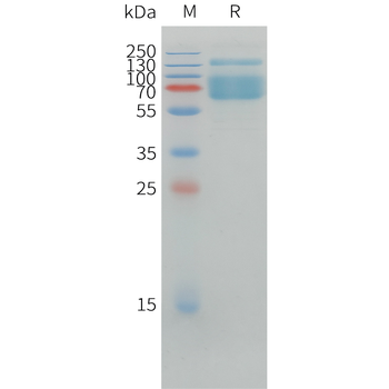
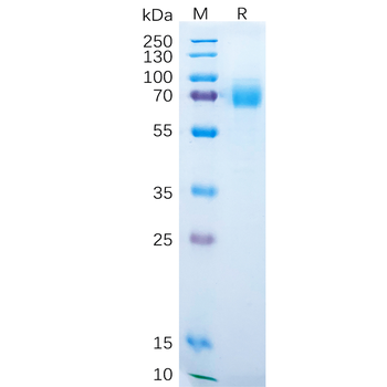
The purity of the protein is greater than 95% as determined by SDS-PAGE and Coomassie blue staining.
The protein has a predicted molecular mass of 36.8 kDa after removal of the signal peptide. The apparent molecular mass of ACVRL1-hFc is approximately 35-70 kDa due to glycosylation.
E.coli
100 μg, 10 μg, 50 μg - FeaturedItem 1 of 3Featured
Unconjugated
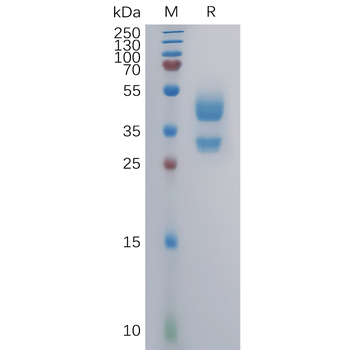
The purity of the protein is greater than 95% as determined by SDS-PAGE and Coomassie blue staining.
The protein has a predicted molecular mass of 10.4 and 37.9 kDa after removal of the signal peptide. The apparent molecular mass of CD3D-His and CD3E-hFc is approximately 35-55 kDa due to glycosylation.
Mammalian
10 μg, 50 μg, 100 μg - FeaturedItem 1 of 3Featured
Unconjugated
The purity of the protein is greater than 95% as determined by SDS-PAGE and Coomassie blue staining.
The protein has a predicted molecular mass of 50.5 kDa after removal of the signal peptide. The apparent molecular mass of hFc-FZD7 is approximately 55-70 kDa due to glycosylation.
Mammalian
10 μg, 50 μg, 100 μg - FeaturedItem 1 of 3Featured
Unconjugated
The purity of the protein is greater than 95% as determined by SDS-PAGE and Coomassie blue staining.
The protein has a predicted molecular mass of 39.4 kDa after removal of the signal peptide. The apparent molecular mass of hFc-S100A9 is approximately 35-55 kDa due to glycosylation.
E.coli
10 μg, 50 μg, 100 μg - FeaturedItem 1 of 4Featured
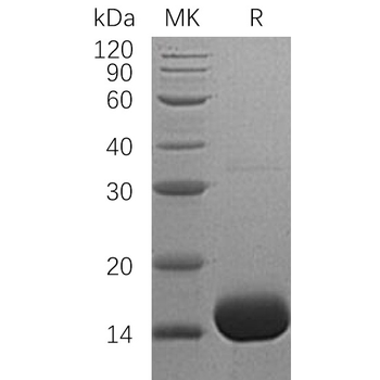
In vitro, In vivo, SDS-PAGE, WB
>95%
~15.1 kDa
Recombinant
100 μg