You have no items in your shopping cart.
Featured
Description
Research Area
Neuroscience
Images & Validation
−Item 1 of 4
| Tested Applications | In vitro, In vivo, WB |
|---|---|
| Application Notes |
Key Properties
−| Source | Synthetic |
|---|---|
| Expression System | Synthetic |
| Biological Origin | Human |
| Target | Amyloid Beta Monomers |
| Reactivity | Human |
| Tag | No Tag |
| Protein Length | 42 amino acids |
| MW | 4.5 kDa |
| Purity | >98% |
| Protein Sequence | DAEFRHDSGYEVHHQKLVFFAEDVGSNKGAIIGLMVGGVVIA |
Storage & Handling
−| Storage | -80°C |
|---|---|
| Buffer/Preservatives | Dry powder. See "Other Resources" for re-suspension instructions/protocol. |
| Concentration | N/A - dried peptide film |
| Expiration Date | 6 months from date of receipt. |
| Disclaimer | For research use only |
Alternative Names
−Abeta Protein, Abeta peptide, Amyloid beta peptide, Beta amyloid peptide, amyloid beta precursor protein peptide, APP
Quality Guarantee
Explore bioreagents carefree to elevate your research. All our products are rigorously tested for performance. If a product does not perform as described on its datasheet, our scientific support team will provide expert troubleshooting, a prompt replacement, or a refund. For full details, please see our Terms & Conditions and Buying Guide. Contact us at [email protected].
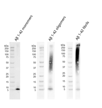
Western blot of amyloid beta 1-42 monomers, oligomers and fibrils using anti-amyloid beta 6E10 antibody. Amyloid beta constructs at 160 pmol were run on 4-12% Bis-Tris SDS-PAGE, transferred to nitrocellulose in the presence of 0.02% v/v Tween-20, and blotted with 1:1000 mouse 6E10 primary antibody (Biolegend). Oligomers observed under TEM/AFM show distinct dimer/trimer bands as well as a signal from ~37-75 kDa (middle). Fibrils observed under TEM/AFM show a signal greater than 100 kDa and a distinct signal in the stacking gel (right).
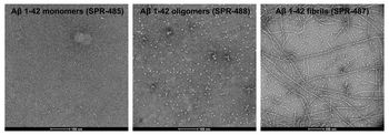
TEM of amyloid beta 1-42 monomers, oligomers and fibrils . Negative stain transmission electron microscopy images acquired at 80 Kv on carbon coated 400 mesh copper grids using phosphotungstic acid and uranyl acetate stain. Scale bar = 100 nm.
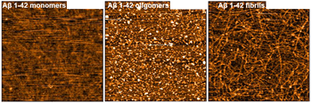
AFM of amyloid beta 1-42 monomers, oligomers and fibrils . Atomic force microscopy analysis of 1.0 mg/mL samples diluted to 0.1 mg/mL in dH2O, mounted on freshly cleaved mica, washed, dried and analyzed with tapping mode. Representative images are 2.5 x 2.5 μm x-y with a z-range of 10 nm.
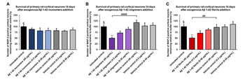
Amyloid beta 1-42 oligomers and fibrils show a dose-dependent toxicity to primary rat cortical neurons, but not monomers. Survival of rat primary cortical neurons 14 days after treatment with different concentrations of (A) monomers, (B) oligomers or (C) fibrils quantified by MAP2 positive neurons and expressed as a percentage of control. Fibrils and respective vehicle controls were initially sonicated in a Bioruptor. Test conditions were run in the same plate as untreated control and vehicle controls, which consisted of buffer without amyloid beta 1-42 protein. Data expressed as mean +/- s.e.m. (n=6).
Quick Database Links
Gene Symbol
Amyloid Beta Monomers
UniProt
UniProt Details
− No UniProt data available
Documents Download
Datasheet
Product Information
Request a Document
Protocol Information
Protein Handling and Storage Guide
Protein Handling Guide
WB
Western Blot (IB, immunoblot)
Amyloid Beta Peptide 1-42 Monomers (orb1714194)

- 0.0
Based on 0 reviews
Participating in our Biorbyt product reviews program enables you to support fellow scientists by sharing your firsthand experience with our products.
Login to Submit a ReviewAvailable Sizes
Select a size below