You have no items in your shopping cart.
Featured
Description
Research Area
Cancer Biology, Cardiovascular Research, Immunology & Inflammation, Metabolism Research
Images & Validation
−Item 1 of 5
| Tested Applications | FC, IF, IHC-P, WB |
|---|---|
| Dilution Range | IF - 1:25, WB - 1:1000, IHC-P - 1:25, FC - 1:25 |
| Reactivity | Human, Mouse |
Key Properties
−| Host | Mouse |
|---|---|
| Clonality | Monoclonal |
| Isotype | IgG1,κ |
| Clone No. | B3553EV016X308X365 |
| Immunogen | Recombinant Protein |
| Target | PPARA |
| Molecular Weight | 52225 |
| Conjugation | Unconjugated |
Storage & Handling
−| Storage | Maintain refrigerated at 2-8°C for up to 2 weeks. For long term storage store at -20°C in small aliquots to prevent freeze-thaw cycles |
|---|---|
| Form/Appearance | Purified monoclonal antibody supplied in PBS with 0.09% (W/V) sodium azide. This antibody is purified through a protein G column, followed by dialysis against PBS. |
| Expiration Date | 12 months from date of receipt. |
| Disclaimer | For research use only |
Alternative Names
−anti Peroxisome proliferator-activated receptor alpha antibody, anti PPAR-alpha antibody, anti Nuclear receptor subfamily 1 group C member 1 antibody, anti PPARA antibody, anti NR1C1 antibody, anti PPAR antibody
Similar Products
−- Item 1 of 7
PPAR alpha Rabbit Polyclonal Antibody [orb15031]
FC, IF, IHC-Fr, IHC-P, WB
Bovine, Equine, Gallus, Porcine, Rabbit
Human, Mouse, Rat
Rabbit
Polyclonal
Unconjugated
50 μl, 100 μl, 200 μl - Item 1 of 9
- Item 1 of 9
- Item 1 of 4
PPAR alpha Rabbit Polyclonal Antibody [orb499655]
FC, IF, IHC-Fr, IHC-P, WB
Canine, Equine, Gallus, Porcine, Rabbit, Rat, Sheep
Human, Mouse
Rabbit
Polyclonal
Unconjugated
50 μl, 100 μl, 200 μl - Item 1 of 7
PPAR-α Polyclonal Antibody [orb1412440]
IF, IHC-P, WB
Human, Mouse, Rat
Rabbit
Polyclonal
Unconjugated
100 μl


Quality Guarantee
Explore bioreagents carefree to elevate your research. All our products are rigorously tested for performance. If a product does not perform as described on its datasheet, our scientific support team will provide expert troubleshooting, a prompt replacement, or a refund. For full details, please see our Terms & Conditions and Buying Guide. Contact us at [email protected].
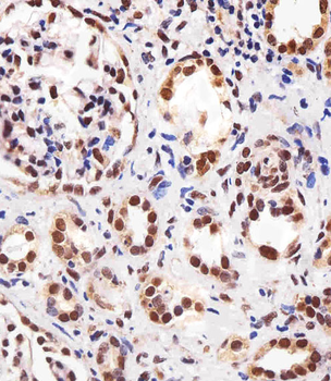
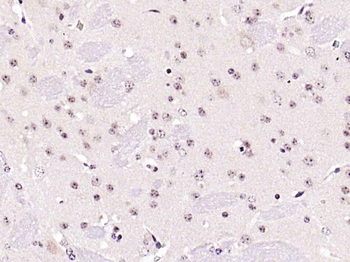
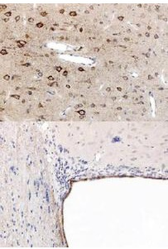
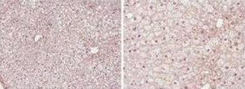
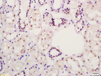
Immunohistochemical analysis of paraffin-embedded H. kidney section using PPARA Antibody. Diluted at 1:25 dilution. A peroxidase-conjugated goat anti-mouse IgG at 1:400 dilution was used as the secondary antibody, followed by DAB staining.
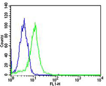
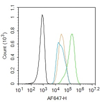
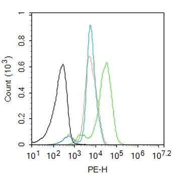
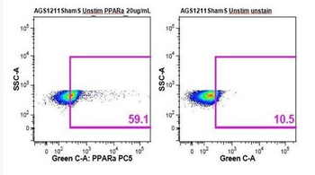
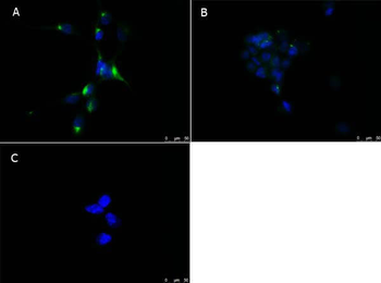
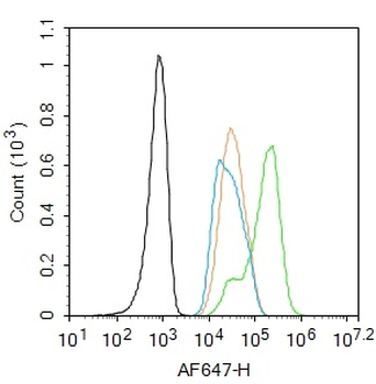
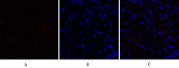
Flow cytometric analysis of Hela cells using PPARA Antibody (green) compared to an isotype control of mouse IgG1 (blue). Diluted at 1:25 dilution. An Alexa Fluor 488 goat anti-mouse lgG at 1:400 dilution was used as the secondary antibody.
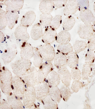
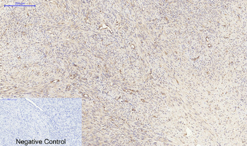
Immunohistochemical analysis of paraffin-embedded H. skeletal muscle section using PPARA Antibody. Diluted at 1:25 dilution. A peroxidase-conjugated goat anti-mouse IgG at 1:400 dilution was used as the secondary antibody, followed by DAB staining.
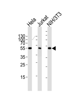
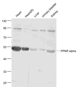
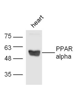
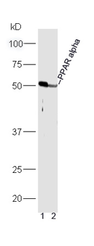
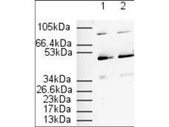
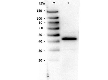
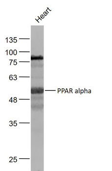
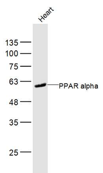
Western blot analysis of lysates from Hela, Jurkat, mouse NIH/3T3 cell line (from left to right), using PPARA Antibody. Diluted at 1:1000 at each lane. A goat anti-mouse IgG H&L (HRP) at 1: 3000 dilution was used as the secondary antibody. Lysates at 35 μg per lane.
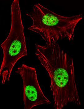
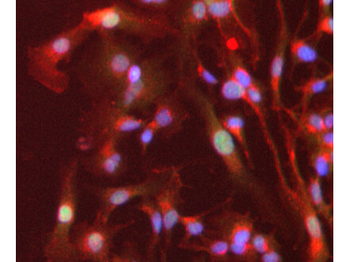
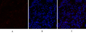
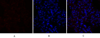
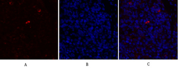
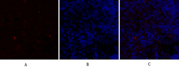
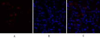
Fluorescent image of Hela cells stained with PPARA Antibody. Diluted at 1:25 dilution. An Alexa Fluor 488-conjugated goat anti-mouse lgG at 1:400 dilution was used as the secondary antibody (green). Cytoplasmic actin was counterstained with Alexa Fluor 555 conjugated with Phalloidin (red).
Quick Database Links
Gene Symbol
PPARA
UniProt
UniProt Details
− No UniProt data available
Documents Download
Datasheet
Product Information
Request a Document
Protocol Information
WB
Western Blot (IB, immunoblot)
IHC-P
Immunohistochemistry Paraffin
FC
Flow Cytometry
IF
Immunofluorescence
PPARA Antibody (orb305792)

- 0.0
Based on 0 reviews
Participating in our Biorbyt product reviews program enables you to support fellow scientists by sharing your firsthand experience with our products.
Login to Submit a ReviewAvailable Sizes
Select a size below
Choose Conjugation or Carrier Free Version
Free Secondary Antibody (20 ul)0/0
Please add an antibody product to your cart first.